Anatomy Heart Valves
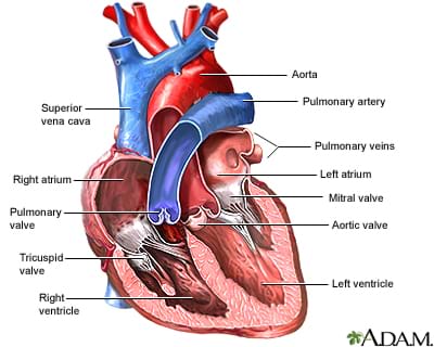
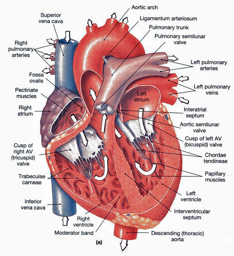
Four valves maintain the unidirectional flow of blood through the heart. Understanding heart valves anatomy is important in grasping the overall function of the heartthe heart is one of the most important organs in the bodyit is responsible for propelling blood to every organ system including itself.
 Pin By Nohemi Daniela On Nursing Cardiac Nursing Nursing
There are four heart valves and.
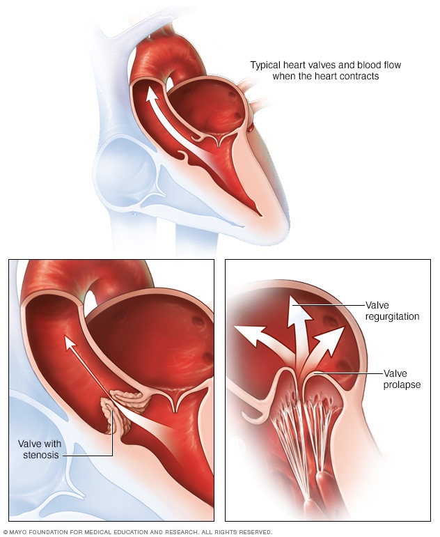
Anatomy heart valves. Blood passes through a valve before leaving each chamber of the heart. A damaged valve forces the heart to re pump blood over and over because the valve doesnt close properly and backflow occurs. Anatomy and function of the heart valves what are heart valves.
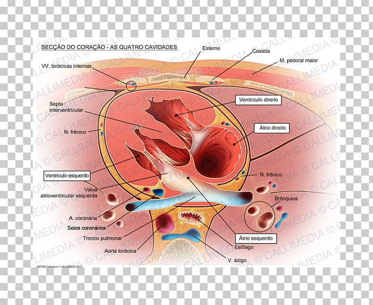
The heart has 4 chambers 2 upper chambers atria and 2 lower chambers ventricles. Heart valves like any mechanical pump can function with leaks but if a valve is badly damaged it can greatly reduce cardiac function. The heart has two kinds of valves atrioventricular and semilunar valves.
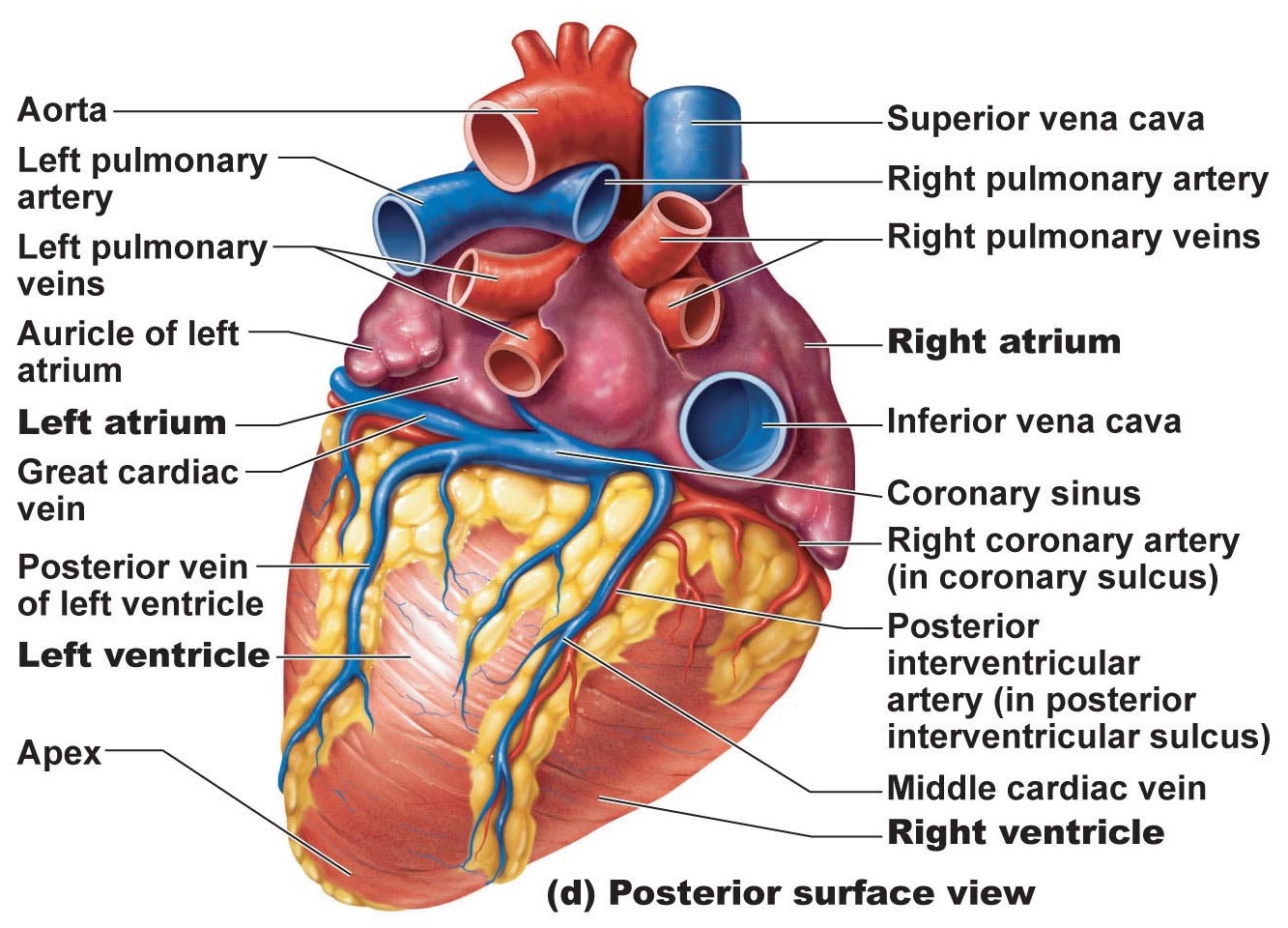
Other articles have discussed at length the gross anatomy of the heart and its four chambers. Introduction to the anatomy of the heart valves. Anatomy of heart valve disease disorders infections.
Heart valves are vital to the proper circulation of blood in the body. Special mention has also been made of the fact that the heart has a. A heart valve opens or closes incumbent on differential blood pressure on each side.
The four valves in the mammalian heart are. The lower two chambers are the right and left ventricles. A heart valve normally allows blood to flow in only one direction through the heartthe four valves are commonly represented in a mammalian heart that determines the pathway of blood flow through the heart.
In this article we will look at the anatomy of these valves their structure function and their clinical correlations by openstax college cc by 30 via wikimedia commons fig 1 the four valves of the heart visible with the atria and great vessels removed. Valves are actually flaps leaflets that act as one way inlets for blood. The upper two chambers are the right and left atria.
The valves prevent the backward flow of blood. These valves open and close during the cardiac cycle to direct the flow of blood through the heart chambers and out to the rest of the body. The valves are located between the atria and ventricles and in the two arteries that empty blood from the ventricles.
Webmds heart anatomy page provides a detailed image of the heart and provides information on heart conditions tests and treatments. Blood is pumped through the chambers aided by four heart valves. Valves are flap like structures that allow blood to flow in one direction.
 Heart Valves Anatomy Mitral Valve Pulmonary Valve Aortic Valve
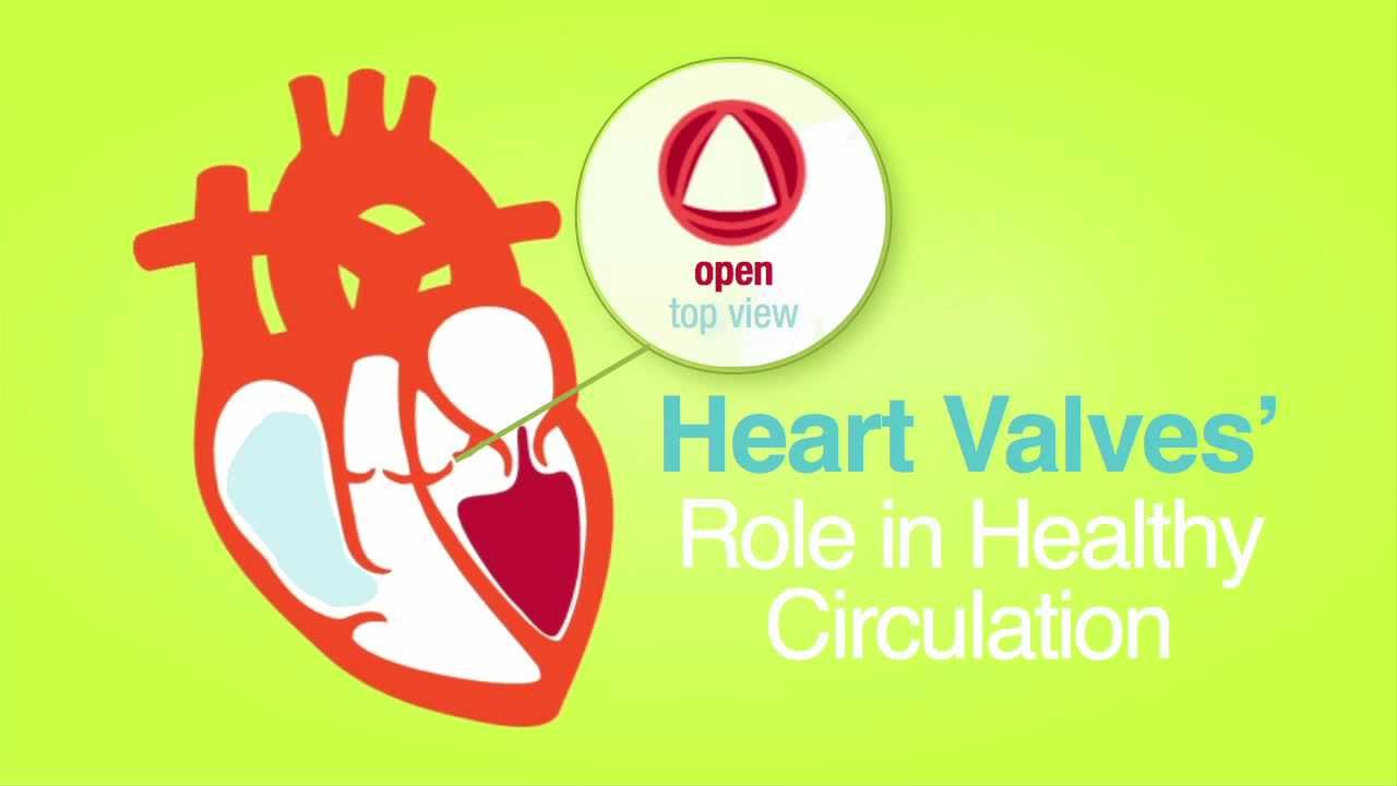 Roles Of Your Four Heart Valves American Heart Association
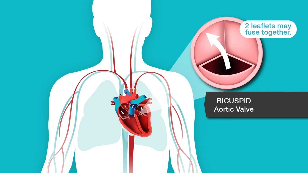 Aortic Stenosis Overview American Heart Association
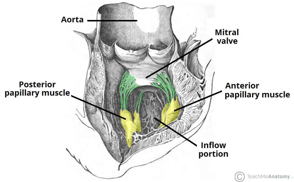 The Heart Valves Tricuspid Aortic Mitral Pulmonary
:watermark(/images/watermark_only.png,0,0,0):watermark(/images/logo_url.png,-10,-10,0):format(jpeg)/images/anatomy_term/valva-atrioventricularis-dextra-valva-tricuspidalis/PjlqmzOPATNgvrEfZp66g_Valva_atrioventriculare_dextra_01.png) Heart Valves Anatomy Tricuspid Aortic Mitral Pulmonary Kenhub
Heart Valves Anatomy Tricuspid Aortic Mitral Pulmonary Kenhub
 What Do I Need To Know About Heart Valves Lesson
 Aortic Stenosis Causes Symptoms And Progression What Is
 A Normal Heart And Heart Valve Problems Mayo Clinic
 Valves Of The Heart Preview Human Anatomy Kenhub
 Heart Anatomy Chambers Valves And Vessels Anatomy
 Heart Anatomy Chambers Valves And Vessels Anatomy
:background_color(FFFFFF):format(jpeg)/images/library/12603/Right_atrium__1_.png) Heart Valves Anatomy Tricuspid Aortic Mitral Pulmonary Kenhub
Heart Valves Anatomy Tricuspid Aortic Mitral Pulmonary Kenhub
 Novel Technique Reduces Obstruction Risk In Heart Valve
:background_color(FFFFFF):format(jpeg)/images/library/11112/left-atrium-and-ventricle_english.jpg) Heart Anatomy Structure Valves Coronary Vessels Kenhub
Heart Anatomy Structure Valves Coronary Vessels Kenhub
 Heart Valve Anatomy Tricuspid Valve Heart Png Clipart
Mitral Valve Annulus Anatomy Structure Pictures
 Heart Valve Right Atrium Aorta Anatomy Png Clipart Anatomy
Heart Valve Flaps What Should Patients Know
 An Overview Of Heart Valve Disease
 Heart Valves Heart Valves Physiology Anatomy
 Heart Valves Showing Pulmonary Valve Mitral Valve And Tricuspid Canvas Print
 Heart Valve Disease Frankel Cardiovascular Center


Belum ada Komentar untuk "Anatomy Heart Valves"
Posting Komentar