Chest Area Anatomy
A mans chest like the rest of his body is covered with skin that has two layers. It contains skin muscle and fatty tissues.
The thorax or chest is a part of the anatomy of humans and various other animals located between the neck and the abdomen.

Chest area anatomy. The muscular makeup of the chest wall includes the intercostal transverses pectoralis. Use the mouse scroll wheel to move the images up and down alternatively use the tiny arrows on both side of the image to move the images. The pelvis is made up of several bones and is at the base of your trunk.
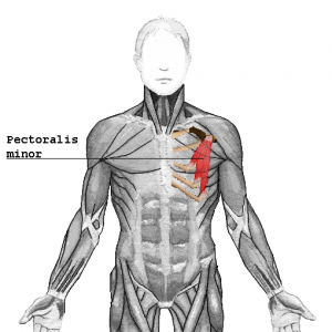
The epidermis is the outermost layer that provides a protective waterproof seal over the body. Helpful trusted answers from doctors. The chest anatomy includes the pectoralis major pectoralis minor and the serratus anterior.
This mri chest thorax axial cross sectional anatomy tool is absolutely free to use. The thorax includes the thoracic cavity and the thoracic wall. The dermis is the under layer that contains sweat glands hair follicles blood vessels and more.
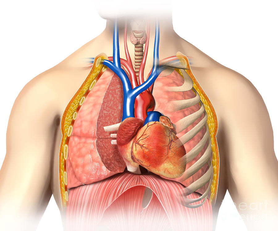
Human female cardiovascular system 7 photos of the human female cardiovascular system human anatomy cardiovascular system human body cardiovascular system human cardiovascular system images human cardiovascular system is considered closed because human cardiovascular system lab report human anatomy human. The circulatory system does most of its work inside the chest. Related posts of anatomy of the chest area human female cardiovascular system.
It contains organs including the heart lungs and thymus gland as well as muscles and various other internal structures. The chest is the area of origin for many of the bodys systems as it houses organs such as the heart esophagus trachea lungs and thoracic diaphragm. The skeletal makeup of the chest wall includes the thoracic vertebrae the ribs and the sternum.
This page provides an overview of the chest muscle group. Fein on human anatomy chest area. It holds your abdominal organs inside houses the genitals and articulates with the thighs and base of spine.
There is no pelvic bone. Learn about each of these muscles their locations functional anatomy and exercises for them. Anatomy of the chest wall.
It is also detrimental in the movement of the upper arms. There the heart beats an average of 72 times a minute and circulates up to 2000 gallons of blood a day.
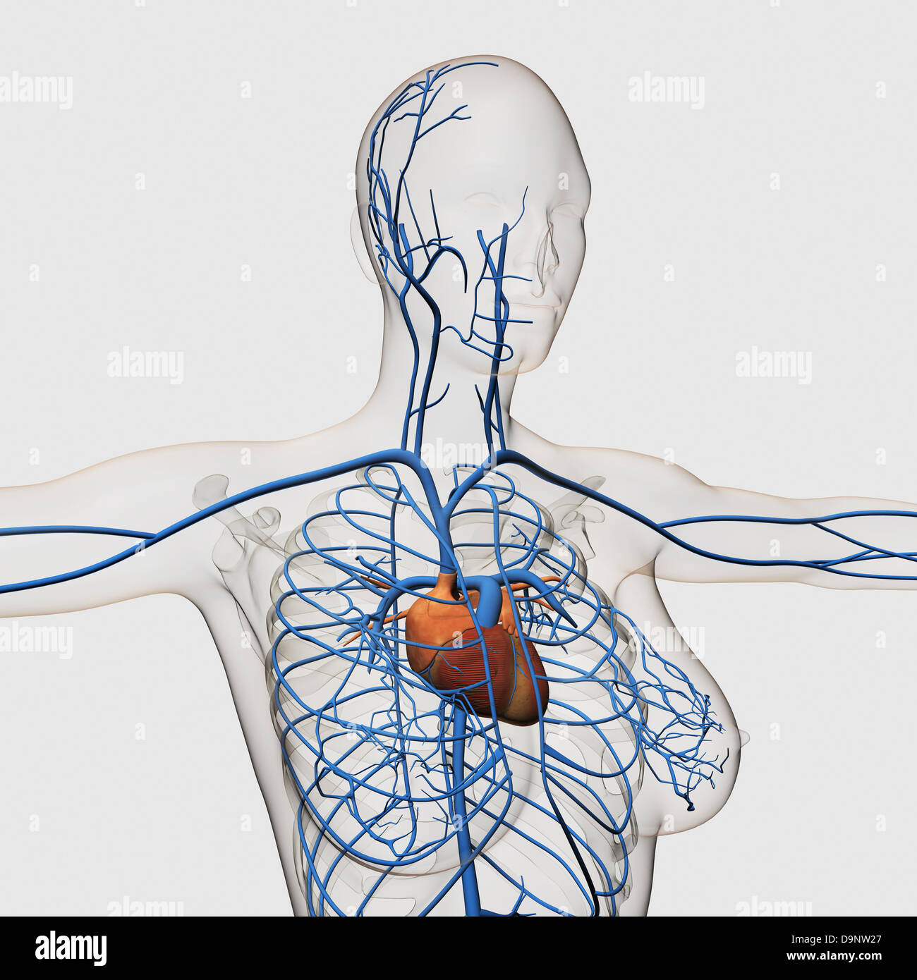 Medical Illustration Of Circulatory System With Heart And
 Chest Wall Tumors The Patient Guide To Heart Lung And
 Understanding The Human Chest Thorax Health Life Media
 Male Anterior Thoracic Wall Chest Iphone X Case
 Massage Therapy For Upper Back Pain
 Muscles In Chest Area Human Chest Muscles Pectoral Muscles
When We Are Doing Cpr Why Do We Compress The Sternum Area
 Cpr In Adults Positioning Your Hands For Chest Compressions
 Medical Illustration Of Arteries Veins And Lymphatic System
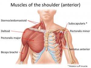 The Neglected Role Of The Chest Muscles In Singing
 Thoracic Oncology Program Malignant Mesothelioma


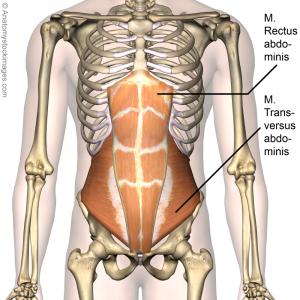 Sternal Pain Different Causes Physiopedia
 Muscles Of The Abdomen Lower Back And Pelvis
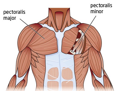 Tight Chest Muscles Why Your Upper Back Is The Key To Their
 Lateral Anatomy Of The Chest Bones And Abdomen Medical
 Human Respiratory System The Mechanics Of Breathing
 Pectoral Muscles Area Innervation Function Human




Belum ada Komentar untuk "Chest Area Anatomy"
Posting Komentar