Anatomy Of The Knee Images
The range of motion of the knee is limited by the anatomy of the bones and ligaments but allows around 120 degrees of flexion. Webmds knee anatomy page provides a detailed image and definition of the knee and its parts including ligaments bones and muscles.
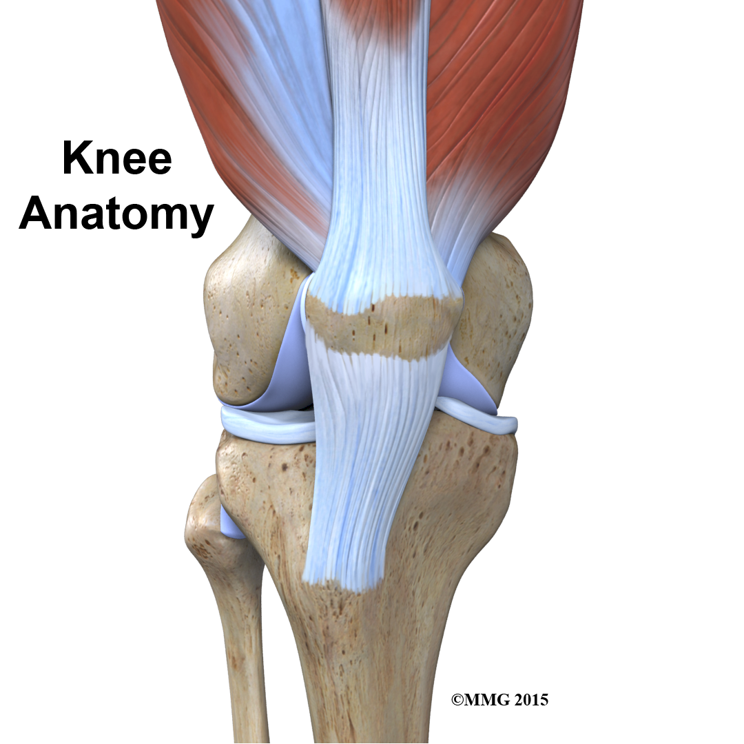
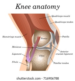
 Knee Anatomy
Click to view large image.
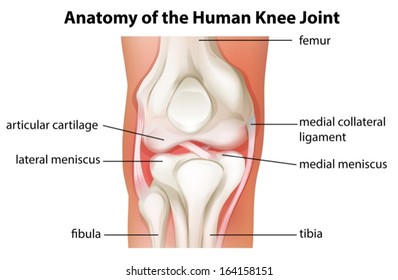
Anatomy of the knee images. Find knee anatomy stock images in hd and millions of other royalty free stock photos illustrations and vectors in the shutterstock collection. Explore basic knee and acl anatomy. The knee joint is made up of three bones and a variety of ligaments.
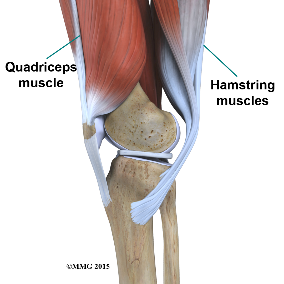
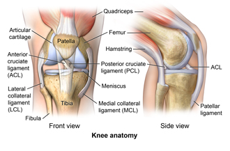
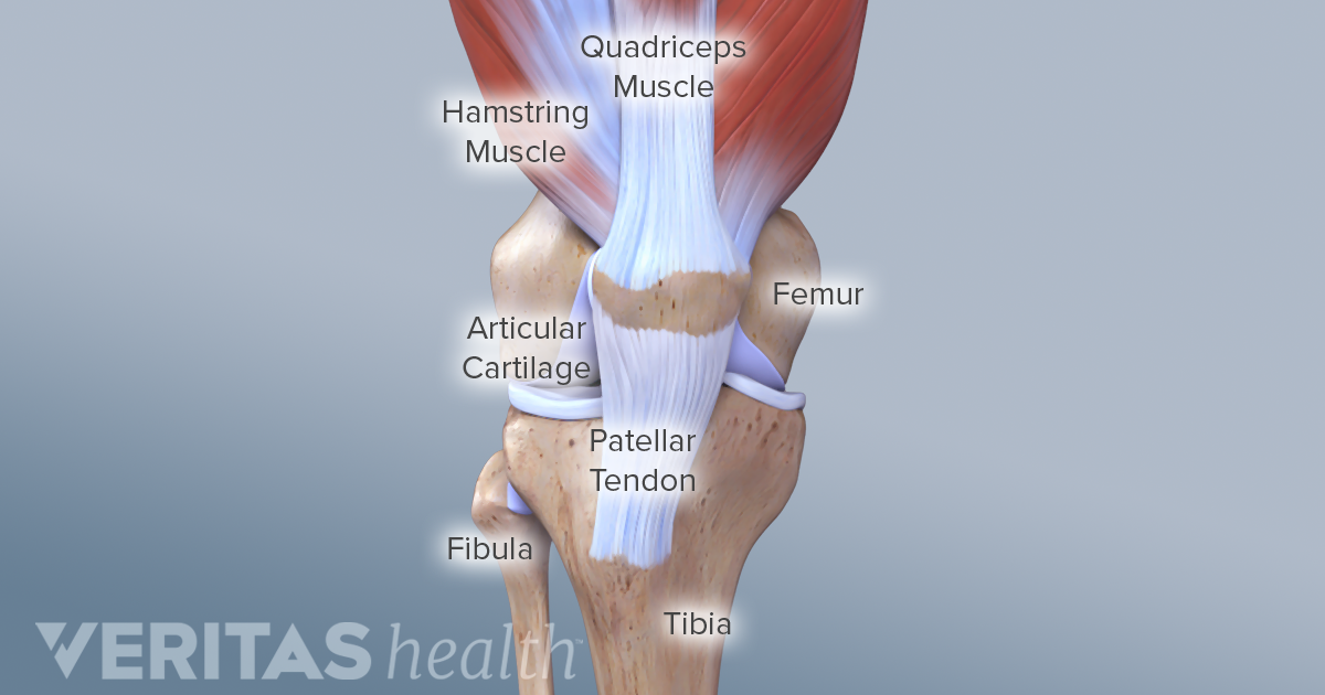
Tendons at the knee. The main tendon found at the knee is the patellar tendon which links the quads muscles to the shin bone. Affordable and search from millions of royalty free images photos and vectors.
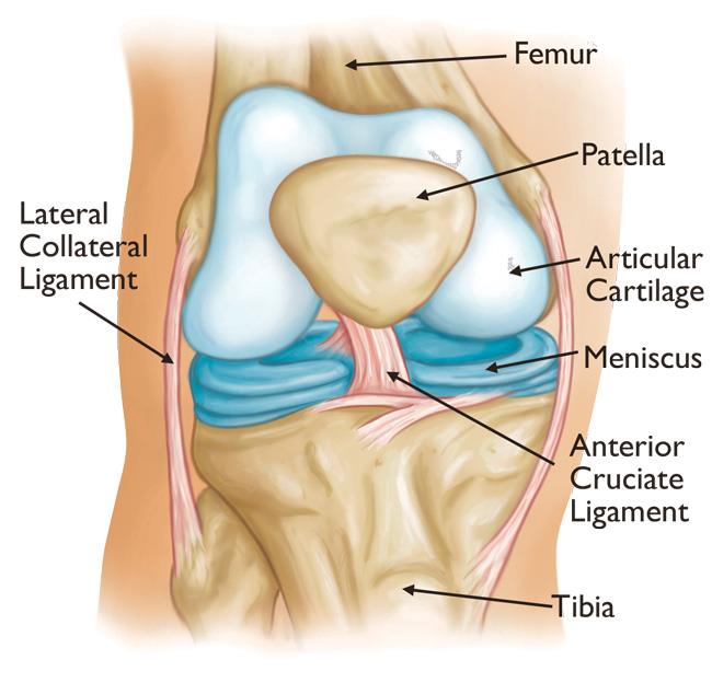
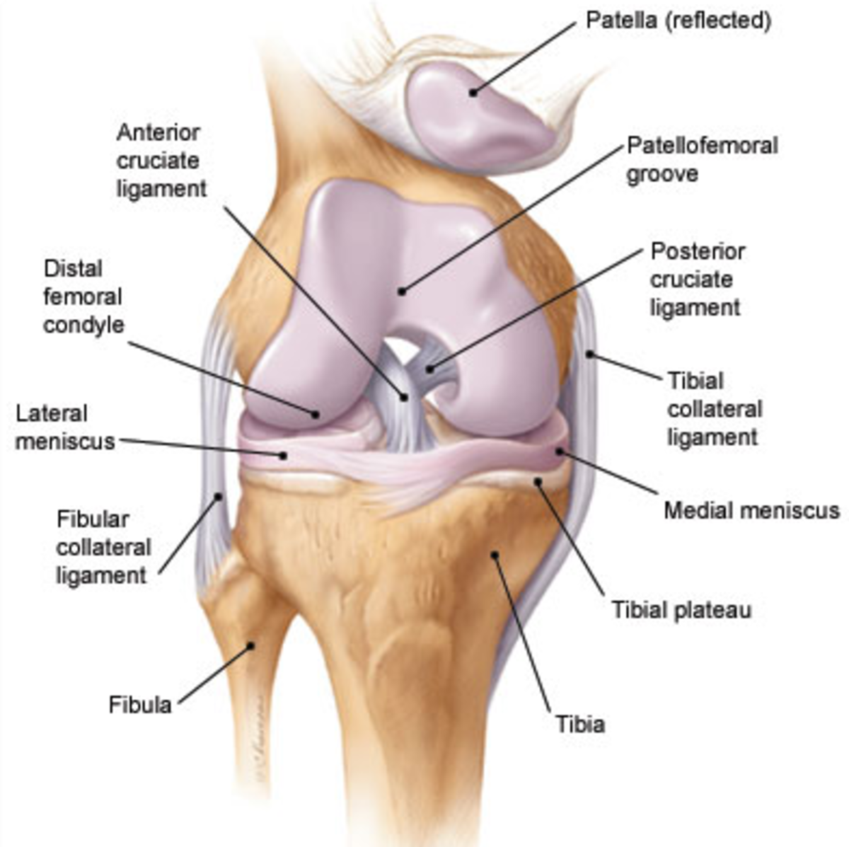
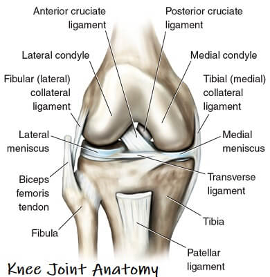
Download knee anatomy stock photos. Between the articular cartilage layer is a shock absorbing cushion called meniscus cartilage. The knee is formed by the femur the thigh bone the tibia the shin bone and the patella the kneecap.
The patella protects the front of the knee joint. The knee cap actually sits inside the patellar tendon. The knee is a complex joint that flexes extends and twists slightly from side to side.
A special characteristic of the knee that differentiates it from other hinge joints is that it allows a small degree of medial and lateral rotation when it is moderately flexed. They are they soft tissues found at the end of muscles which link the muscle to bone. Lets begin with the basics of knee anatomy.
Tendons are often overlooked as part of knee joint anatomy. See the pictures and anatomy description of knee joint bones cartilage ligaments muscle and tendons with resources for knee problems injuries. Thousands of new high quality pictures added every day.
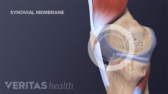
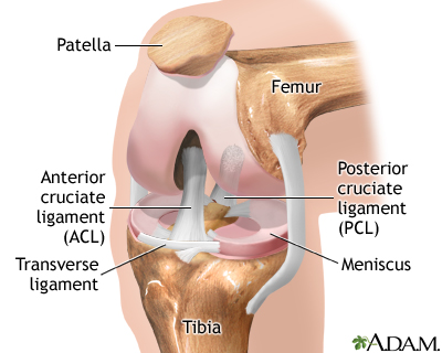
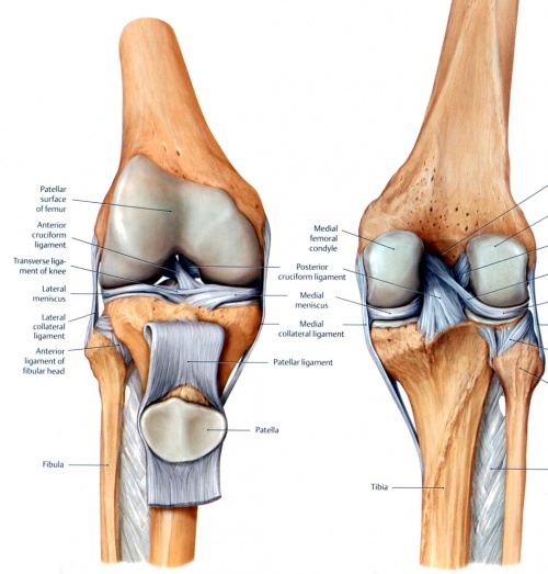
We have a series of diagram images showing the components and function of the knee joint. The knee is the meeting point of the femur thigh bone in the upper leg and the tibia shinbone in the. Knee anatomy springer medizin getty images inside the knee joint is a smooth cover on the ends of the bone called articular cartilage.
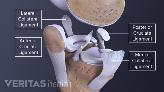
The collateral ligaments run along the sides of the knee and limit the sideways motion of the knee. The knee joint is surrounded by a joint capsule with ligaments strapping the inside and outside of the joint collateral ligaments as well as crossing within the joint cruciate ligaments.
 Physical Therapy In Buffalo For Knee Anatomy
 Knee Anatomy
 Total Knee Replacement Orthoinfo Aaos
 Anatomy Of The Knee Bones Muscles Arteries Veins Nerves
Knee Anatomy
 Knee Anatomy
/188058334-crop-56aae7425f9b58b7d0091480.jpg) What Is Causing Your Knee Pain
What Is Causing Your Knee Pain
 Knee Arthroscopy Series Normal Anatomy Medlineplus
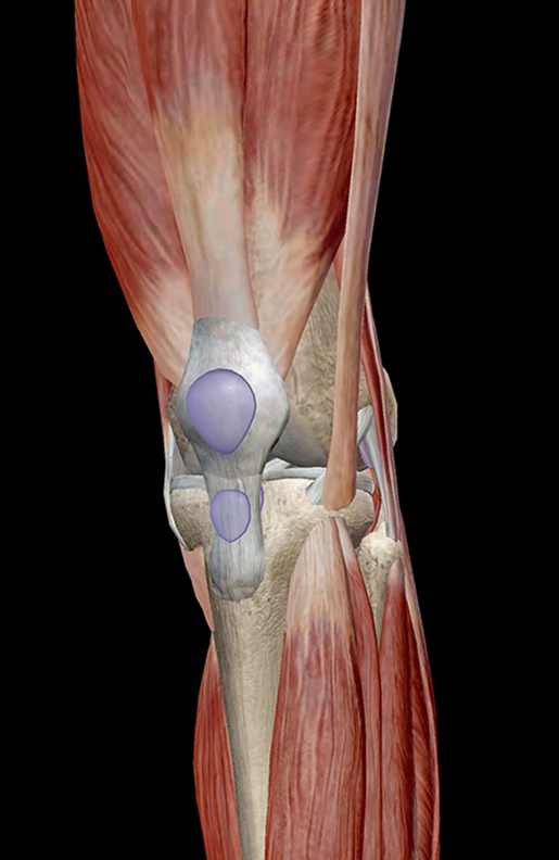
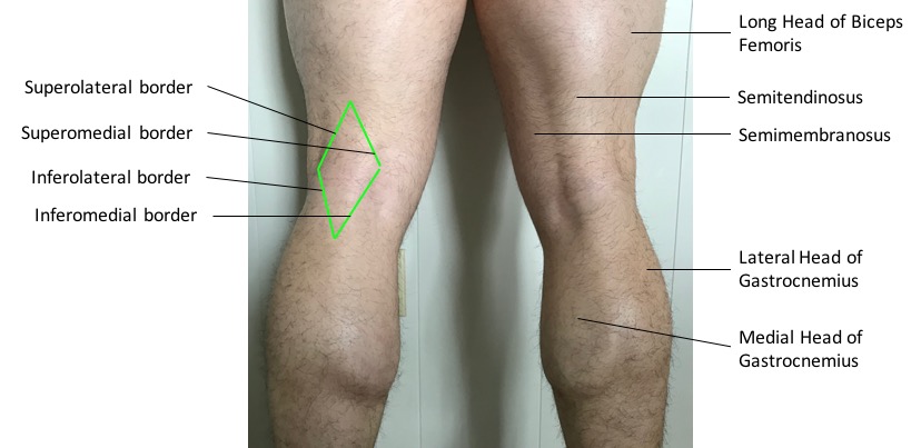 Surface Anatomy Advanced Anatomy 2nd Ed
 Anatomy Knee Joint Klinik Am Ring
 Knee Anatomy Including Ligaments Cartilage And Meniscus
 Knee Joint Anatomy Motion Knee Pain Explained
 Anatomy Of The Knee Joint
 Knee Wikipedia
 2 Anatomy Of Knee Joint Adapted From 34 Download
 Learn Muscle Anatomy Knee Joint Group
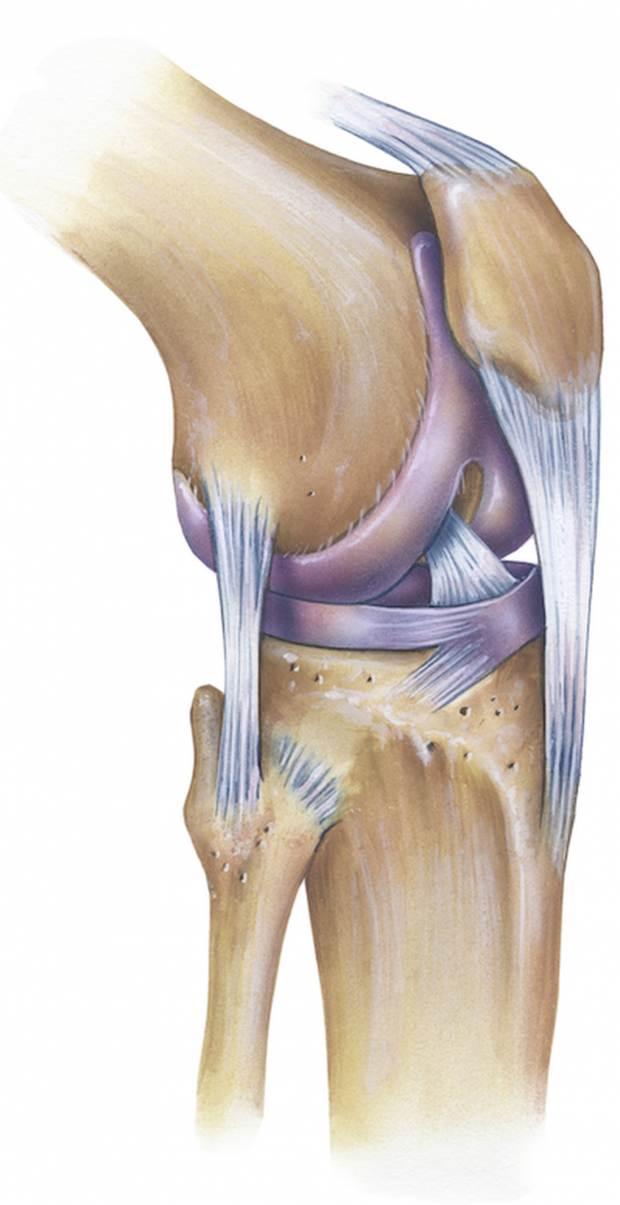
 Knee Joint Anatomy Lateral View
 Knee Physiopedia
 Cicitop Anatomy Science Medical Knee Joint With Ligaments
 Physical Therapy In Buffalo For Knee Anatomy
 Anatomy Subscribe Structure Knee Joint
 Knee Joint Anatomy
 The Human Knee Joint S Anatomy With Visible Cruciate
 Anatomy Of The Knee Central Coast Orthopedic Medical Group
 Knee Anatomy

 Redding Hospital Knee Anatomy
 Knee Anatomy Images Stock Photos Vectors Shutterstock
 Matthew Boyle Orthopaedic Surgeon Knee Anatomy Knee
 Knee Anatomy Images Stock Photos Vectors Shutterstock
 Anatomy Of The Knee Baxter Regional Medical Center
![]() Leg And Knee Anatomy Bones Muscles Soft Tissues Kenhub
Leg And Knee Anatomy Bones Muscles Soft Tissues Kenhub
Belum ada Komentar untuk "Anatomy Of The Knee Images"
Posting Komentar