Basic Eye Anatomy
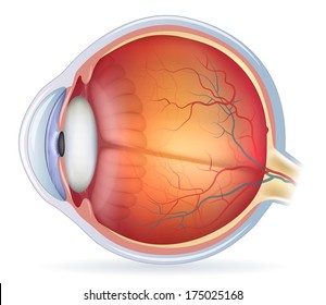
To understand the diseases and conditions that can affect the eye it helps to understand basic eye anatomy. The front sixth of this layer is clear and is called the cornea.
 Basic Eye Anatomy Potthoff Eye Care And Surgery
Six extraocular muscles in the orbit are attached to the eye.

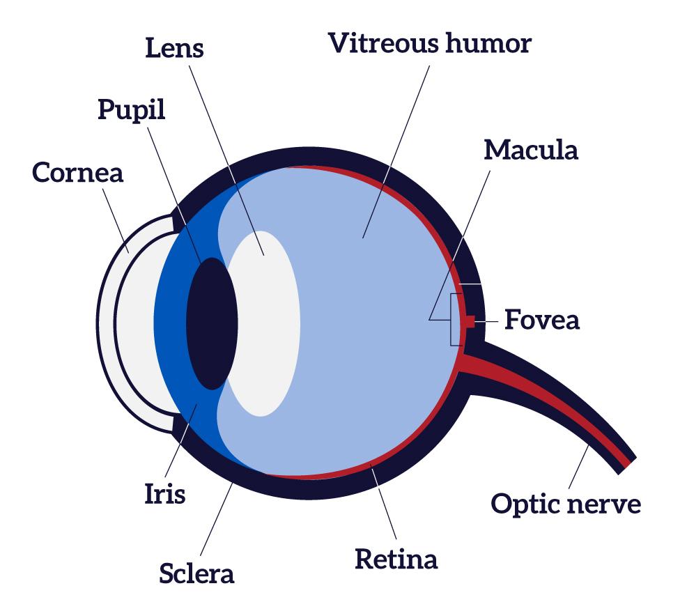
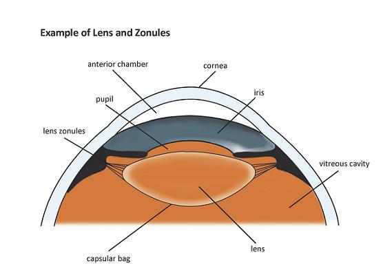
Basic eye anatomy. The eyes crystalline lens is located directly behind the. This is the part of the eye that gives it color ie. The iris of the eye functions like the diaphragm of a camera controlling the amount.
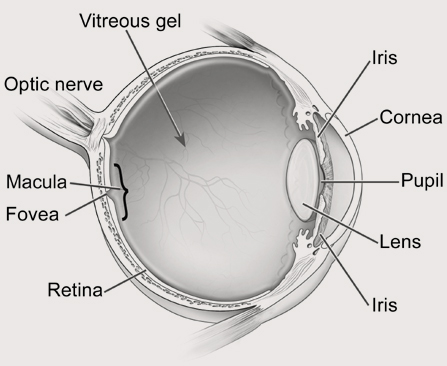
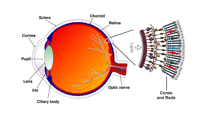
The tough outermost layer of the eye is called the sclera. This is a. A small area in the retina that contains special light sensitive cells.
The crystal clear dome that covers the front of the eye. The eye is approximately 1 inch 254 cm wide 1 inch deep and 09 inches 23 cm tall. The cornea transmits and focuses light into the eye.
Basic eye anatomy cornea. In a number of ways the human eye works much like a digital camera. These muscles move the eye up and down and side to side and rotate the eye.
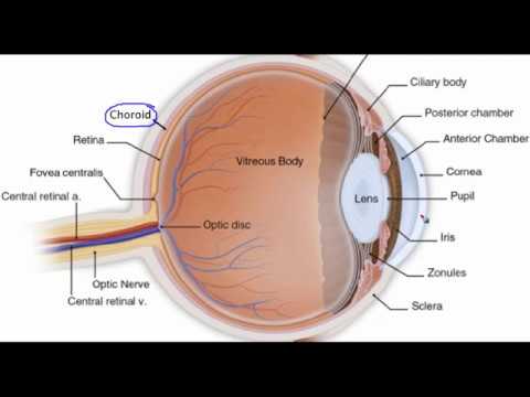
Here is a tour of the eye starting from the outside going in through the front and working to the back. The transparent structure inside the eye that focuses light rays onto the retina. Although small in size the eye is a very complex organ.
The crystalline lens finishes the focusing of light. Basic eye anatomy understanding the anatomy of the eye is critical to understanding cataracts how and why cataracts affect vision and cataract surgery itself. Light is focused primarily by the cornea the clear front surface of the eye.
Although the eye is small relative to most organs in the human body it has many distinct anatomical parts all of which contribute to the production of normal vision in one way or another. It maintains the shape of the eye. Basic eye anatomy.
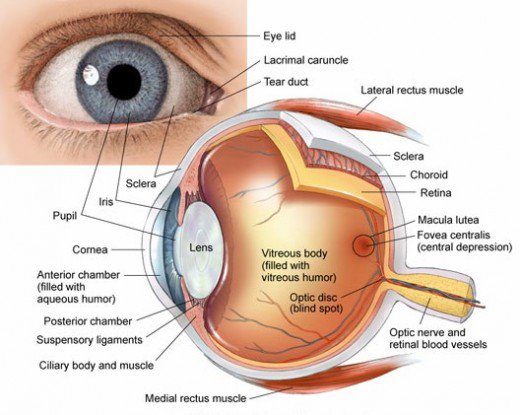
The colored part of the eye. The iris helps regulate the amount of light that enters the eye. This is the opening in the middle of the iris.
The eye sits in a protective bony socket called the orbit.
 Anatomy Of The Eye Ophthalmology Patient Education Eanw
 Anatomy Eye Images Stock Photos Vectors Shutterstock
 Comfortable Face Down Recovery After Eye Surgery Body

 Human Eye Anatomy Structure And Function
 The Basic Anatomy Of A Human Eye Printed With Permission
 How To Draw Human Eye Diagram Step By Step For Beginners
Anatomy Of The Eye 101 Eyecheck
Parts Of The Eye American Academy Of Ophthalmology
 How To Draw Comics How To Draw Eyes
 How The Eyes Work National Eye Institute
 Schematic Representation Of The Basic Anatomy Of The Human
 Eye Anatomy

 C Basic Eye Anatomy Physics Project
 Human Eye Ball Anatomy Physiology Diagram
 2 Minute Topic Basic Anatomy Of The Human Eye
 Human Eye Anatomy Parts And Structure Online Biology Notes
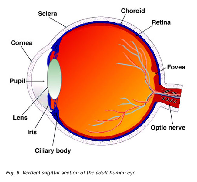
 Figure 3 1 Basic Anatomy Of The Eye Fovea Is The Area Of
 Anatomy Of The Eye Retina Centers
 Deylah S Muffiny Anatomy Blog Basic Eye Anatomy Worksheet
 Eye Anatomy A Closer Look At The Parts Of The Eye
 Pin On Ophthalmology
 How The Eyes Work Ophthalmologist Eye Surgery Raleigh

 Eye Anatomy And Function
 Anatomy And Structure Of The Eye Brightfocus Foundation
Belum ada Komentar untuk "Basic Eye Anatomy"
Posting Komentar