Anterior Hip Anatomy
Using the femoral head as a landmark the anterior synovial recess is identified. Direct anterior hip replacement is a minimally invasive surgical technique.
The movements of the hip joint is thus performed by a series of muscles which are here presented in order of importance with the range of motion from the neutral zero degree position indicated.

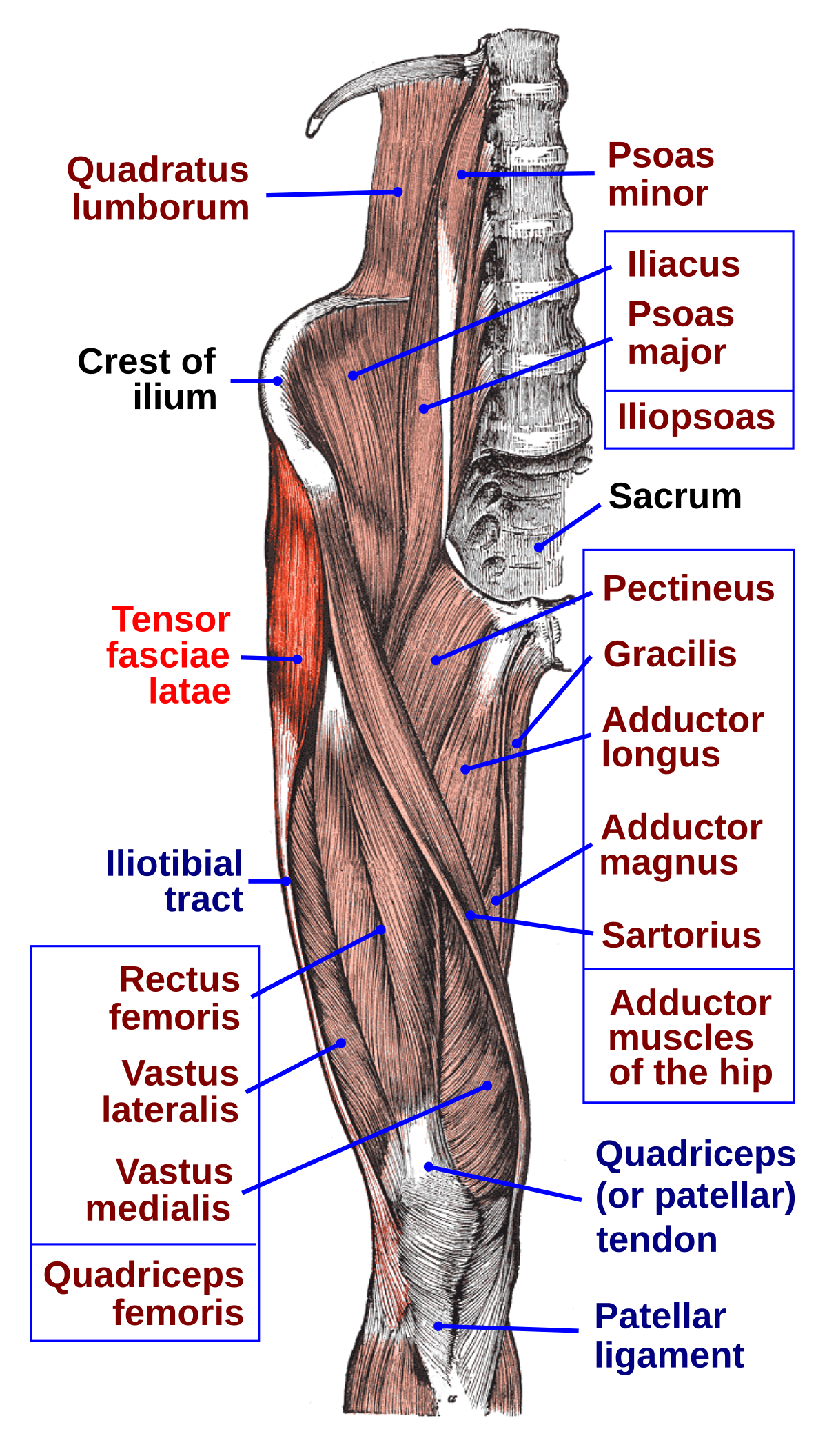
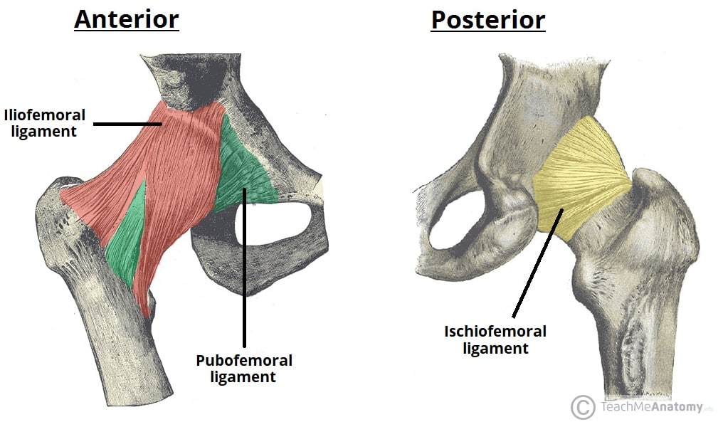
Anterior hip anatomy. The stability of the hip is increased by the strong ligaments that encircle the hip. Retract rectus femoris and iliopsoas medially and gluteus medius laterally to expose the hip capsule. The approach continues between the sartorius and tensor fascia lata muscles an internervous plane avoiding the risk of denervation of the involved muscles.
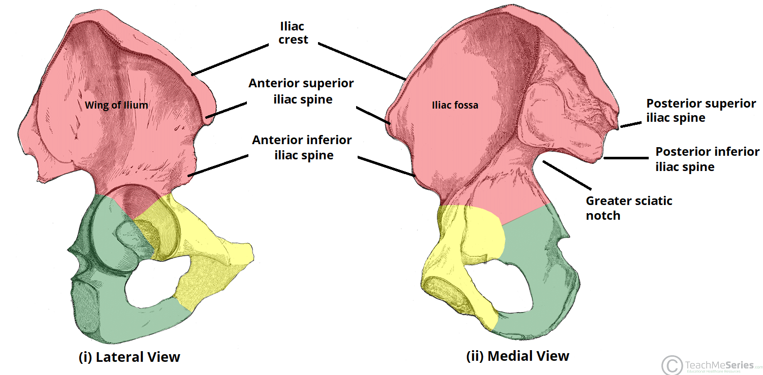
Adduct and externally rotate the hip to place the capsule on stretch. Detach rectus femoris from both its origins. The adult os coxae or hip bone is formed by the fusion of the ilium the ischium and the pubis which occurs by the end of the teenage years.
Muscles of the hip. The hip articulation is true diarthroidal ball and socket style joint formed from the head of the femur as it articulates with the acetabulum of the pelvis. The natural motion of the hip allows us to walk run swim cycle dance for those of us with rhythm and enjoy our lives as upright bipeds with grace.
In hip replacement surgery the hip can be reached through the back of the hip posterior approach the side of the hip lateral or anterolateral approach the front of the leg anterior approach or through a combination of approaches. The hip joint is a ball and socket type joint. This joint serves as the main connection between the lower extremity and the trunk and typically works in a closed kinematic chain.
Anteroposterior and lateral radiographs of the hip. Study of the anterior hip region. In physiological conditions it is virtual whereas it is distended by anechoic fluid in the presence of joint effusion.
Lateral or external rotation 30 with the hip extended 50 with the hip flexed. When the cartilage in the hip joint starts to deteriorate the implications are profound. As a result today there is a range of surgical approaches being utilized by orthopedic surgeons.
This approach involves a 3 to 4 inch incision on the front of the hip that allows the joint to be replaced by moving muscles aside along their natural tissue planes without detaching any tendons. Anatomy of the hip the hip joint. The 2 hip bones form the bony pelvis.
The hip is a ball and socket joint in the engine room of human locomotion. Illustration of the right hip demonstrates the location and size of the incision for the direct anterior approach. The hip joint is the articulation of the pelvis with the femur which connects the axial skeleton with the lower extremity.
Identify plane between rectus femoris and gluteus medius. The muscles of the thigh and lower back work together to keep the hip stable.
 Psoas Major Part I Hip Flexor Or Lumbar Stabilizer
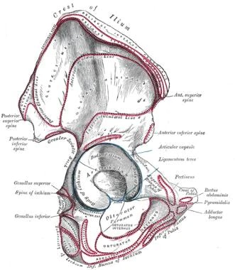 Hip Joint Anatomy Overview Gross Anatomy
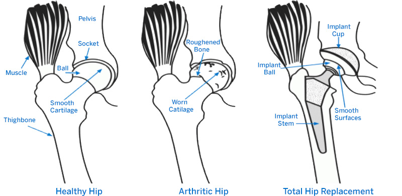 How Do I Know If I Need A Hip Replacement Hss Orthopedics
Basics Of Hip Anatomy Mike Scaduto
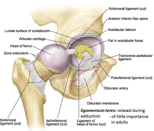 Hip Anatomy Recon Orthobullets
 Muscles Of The Thigh Part 1 Anterior Compartment Anatomy
 Artificial Hip Replacement Anterior Approach
 Muscles Of The Hip Anatomy Pictures And Information
 Anatomy And Injuries Of The Hip Anatomical Chart
 Anterior Right Hip And Thigh Muscle Anatomy Diagram Quizlet
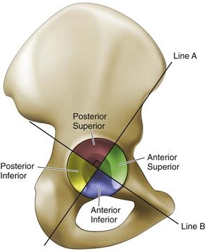 Hip Anatomy Recon Orthobullets
 Tensor Fasciae Latae Muscle Wikipedia
 Pain At The Front Of The Hip Sartorius Psoas Strain
 The Hip Bone Ilium Ischium Pubis Teachmeanatomy
 Hip Adductors Anatomy And Exercises
Hip Pain Orthopedics University Of Colorado Denver
Osteonecrosis Of The Hip Orthoinfo Aaos
 The Hip Joint Articulations Movements Teachmeanatomy
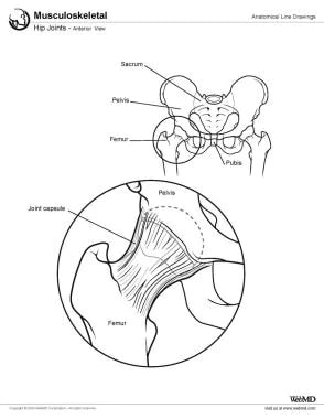 Hip Joint Anatomy Overview Gross Anatomy
 Medacta Corporate Amis A Medacta Solution Why An Amis


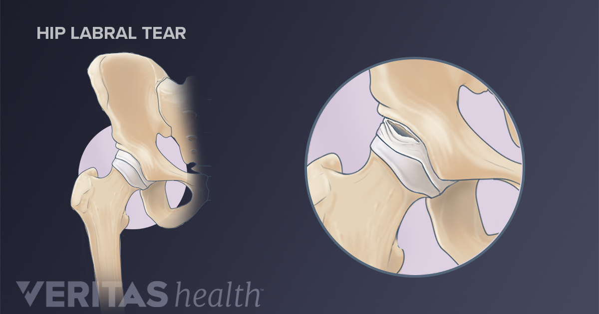

Belum ada Komentar untuk "Anterior Hip Anatomy"
Posting Komentar