Brain Anatomy Ct
Non contrast sagittal ct head. Non contrast axial ct head.
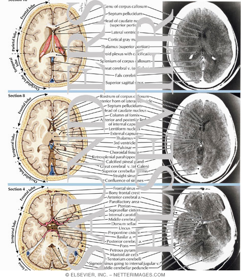
 Brain And Face Ct Interactive Anatomy Atlas
Cross sectionnal anatomy of the head on a cranial ct scan.
Brain anatomy ct. Interactive anatomy atlas. Online mri ct sectional anatomy omcsa k anatomy is probably one of the most user friendly and convenient online interface for human anatomy atlas. Anatomy ct axial brain form no 18.
Non contrast coronal ct head. Brain bones of cranium sinuses of the face. Hnbs neuroanatomy modules neck ct.
Welcome to online mri ct sectional anatomy. Welcome to headneckbrainspine a website intended for those interested in neuroradiology anatomy and learning from neuroradiology cases. This article lists a series of labeled imaging anatomy cases by system and modality.
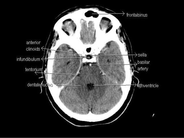
6 frontal bone 27 occipital bone 32 optic nerve 37 basilar artery 40 hemisphere of cerebellum 43 frontal sinus 45 sigmoid sinus 46 internal carotid artery 47 sphenoid bone 49 medulla oblongata 50 external auditory meatus 51 spinal central canal. Jakab m kikinis r. Angiogram coronal ct head.
It contains both regions of gray and white matter in a heterogeneous fashion that are best appreciated on t1 and t2 weighted mr coronal images. See discussion on the basal forebrain in self test question 1. To navigate the website click on the images below or on the above menu.
Brain and face ct. Real time interface human sectional anatomy. Basal forebrain on ct and mr images the basal forebrain is a rather featureless region on the ventral surface of the brain.
Spl head and neck atlas 2012 november. Head and neck atlas. Angiogram axial ct head.
Brain bones of skull paranasal sinuses. Neck ct cervical lymph node levels. Neuroanatomy encompasses the anatomy of all structures of the central nervous system which includes the brain and the spinal cord and their supporting structures.
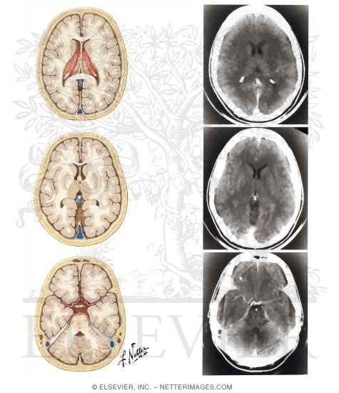
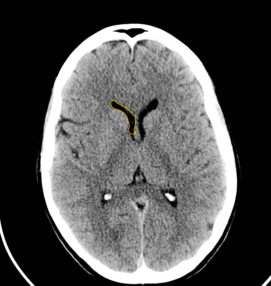
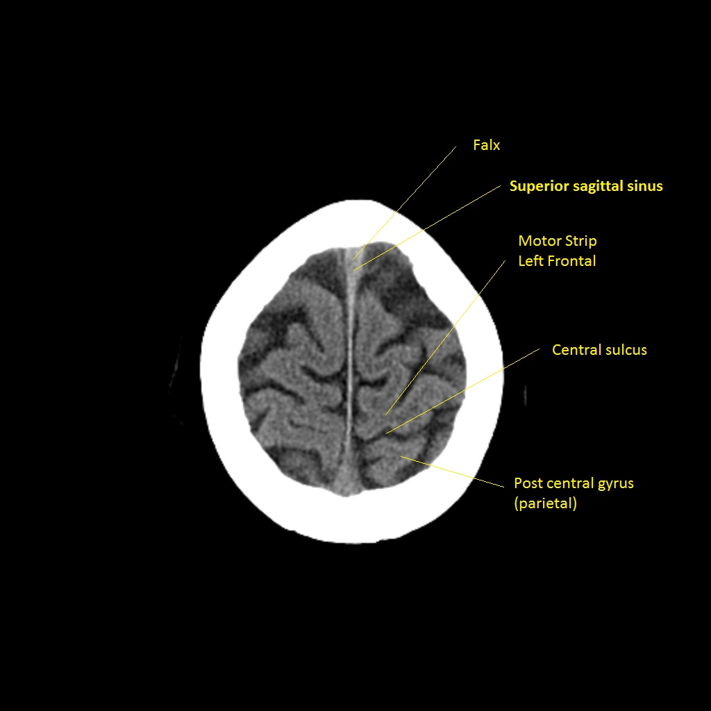
Ct images of the brain are conventionally viewed from below as if looking up into the top of the head. To load the neck ct anatomy module in a new window click on its image above. Coronal brain ct.
This means that the right side of the brain is on the left side of the viewer. Given that the file is large loading may take a few minutes. Ct brain image orientation.
Thoracolumbar junction ct. Anatomy of the head on a cranial ct scan. Ct head neck atlas.
This anatomy section promotes the use of the terminologia anatomica the global standard for correct gross anatomical nomenclature. The anterior part of the head is at the top of the image.
 Section Vi Head Ct Emergency Radiology Case Studies
![]() Medical Imaging And Radiological Anatomy X Ray Ct Mri
Medical Imaging And Radiological Anatomy X Ray Ct Mri
 Normal Brain Anatomy As Demonstrated By Computerized
 Age And Anatomy Related Values Of Blood Brain Barrier
 Brain Lobes Annotated Mri Radiology Case Radiopaedia Org
 Basic Ct Anatomy Of The Brain
 Brain And Face Ct Interactive Anatomy Atlas
 Ct Anatomy
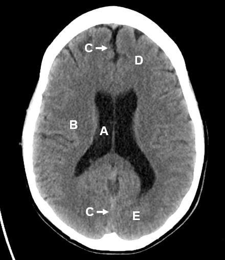
 Normal Ct Brain Radiology Case Radiopaedia Org
 Head Ct
 Ct Scan Hemorrhagic Stroke Stock Image Image Of Hospital
 Learn Ct Scan Anatomy Ct Axial Brain
 Presentation1 Pptx Radiological Anatomy Of The Brain
 Cross Sectional Anatomy Brain And Spinal Column Brain
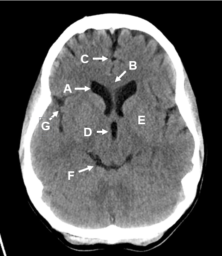
 Ct Head Normal Anatomy
 Normal Brain Anatomy As Demonstrated By Computerized
 Axial View Of A Head Computed Tomography Ct Scan Of Pineal
 The Radiology Assistant Brain Anatomy
 Basic Anatomy Of Ct Brain Hku E Learning Platform In
 Head Ct Scan Procedure Radtechonduty
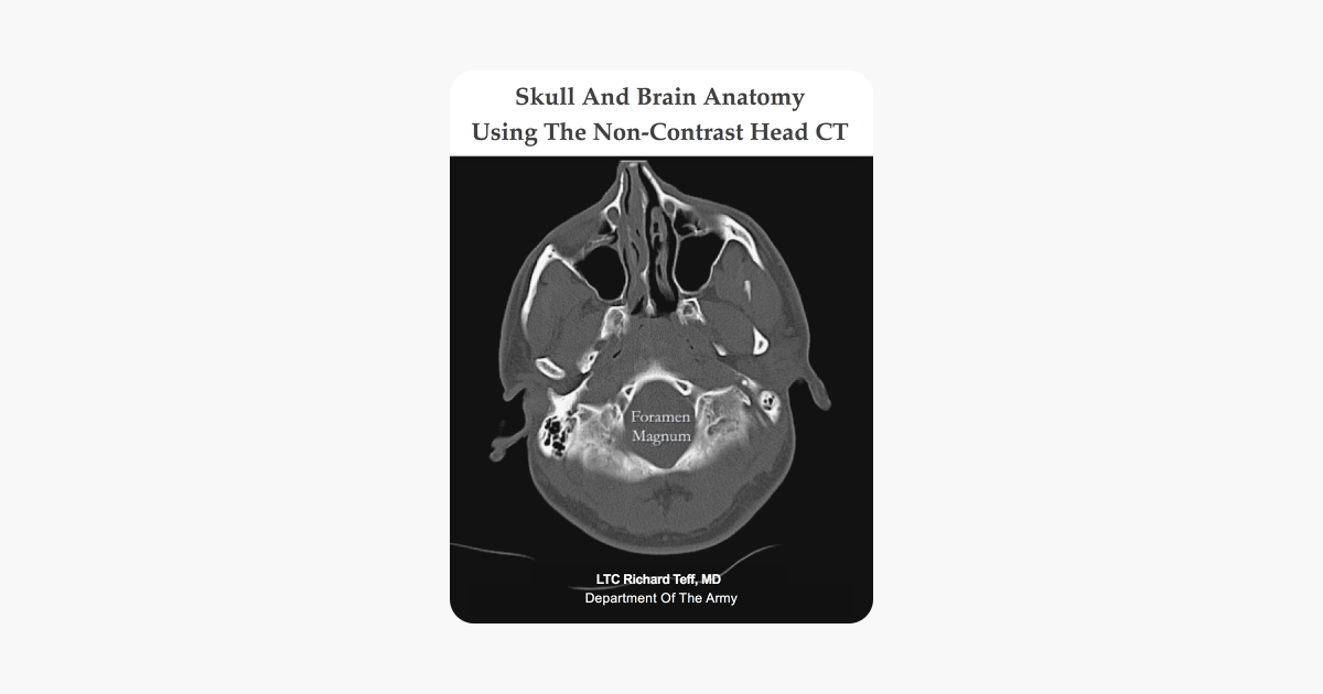 Skull And Brain Anatomy Using The Non Contrast Head Ct
 Brain And Spines Brain Anatomy Ct
 Glioblastoma Brain Cancer Ct Scan License Download Or
 Imaging Of The Central Nervous System Clinical Gate
 Normal Anatomy Of The Brain On Ct And Mri With A Few Normal
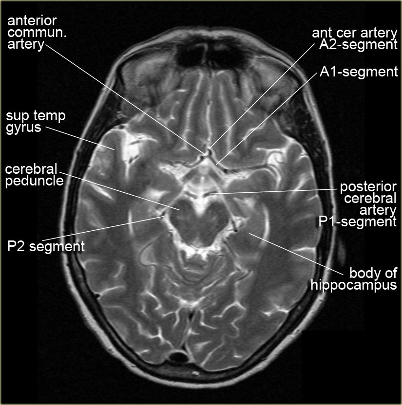
 Mri Anatomy Free Mri Axial Brain Anatomy
 Basic Anatomy Of Ct Brain Hku E Learning Platform In
Belum ada Komentar untuk "Brain Anatomy Ct"
Posting Komentar