Anatomy Of A Vein
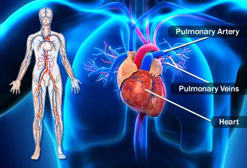
Their anatomy is discussed in the following sections see figure 23 1. The pulmonary veins along with the pulmonary arteries make up the pulmonary circulation.
Anatomy of arteries vs veins.
Anatomy of a vein. The pulmonary veins can be affected by. Because the arteries arterioles and capillaries absorb most of the force of the hearts contractions veins and venules are subjected to very low blood pressures. These are located in the fatty layer under your skin.
Types of veins systemic veins. To ensure unrestricted flow of blood a venule blood vessel allows deoxygenated blood to return from the capillary beds to the vein. Veins are composed of three main layers.
In contrast to veins arteries carry blood away from the heart. They carry the deoxygenated blood which is bluish in color and for the same reason veins appear blue. The vein also contains venous valves one way flaps that prevent blood from flowing back and pooling in the lower extremities due to the effects of gravity.
Their lumen is larger than that of the accompanying arteries. The superficial veins of the upper limb are the veins selected for most elective venipuncture. Deep veins and superficial veins deep veins.
The adventitia fuses with surrounding tissue in the body. There are valves in most veins to prevent backflow. Exceptions are the pulmonary and umbilical veins both of which carry oxygenated blood to the heart.
The pulmonary veins serve a very important purpose of delivering freshly oxygenated blood. The tunica media or media is the middle layer of a veins wall. Veins are blood vessels that carry blood towards the heart.
The anatomy of the pulmonary vein anatomy. A vein is a blood vessel that conducts blood toward the heart. A vein can range in size from 1 millimeter to 1 15 centimeters in diameter.
Veins are the large return vessels of the body and act as the blood return counterparts of arteries. Because they are low pressure vessels larger veins are commonly equipped with valves that promote the unidirectional flow of blood toward the heart and prevent backflow toward the. Veins are the blood vessels which carry the blood from peripheral tissues towards heart.
Blood from superficial veins is often directed into the deep veins through short veins. These are found in muscles or along bones. Compared to arteries veins are thin walled vessels with large and irregular lumens see figure 6.
This layer is built of collagen elastic fibers and smooth muscle fibers. Veins are thin walled being thinner than the arteries. Blood to the digits is drained through an anastomosis of palmar and dorsal digital veins.
Veins are less muscular than arteries and are often closer to the skin. The smallest veins in the body are called venules. This layer is the thickest layer of a veins lining and is made of loose connective tissues and an external elastic membrane.
Most veins carry deoxygenated blood from the tissues back to the heart.
 External Jugular Vein Earth S Lab
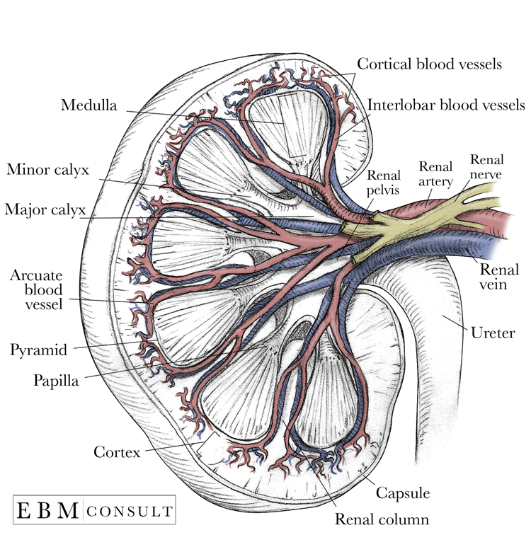 Kidney Anatomy Image
 Venous System
 Human Body Skin Anatomy Vector Illustration With Parts Vein Artery
 Diagram Showing The Venous Anatomy Of The Leg
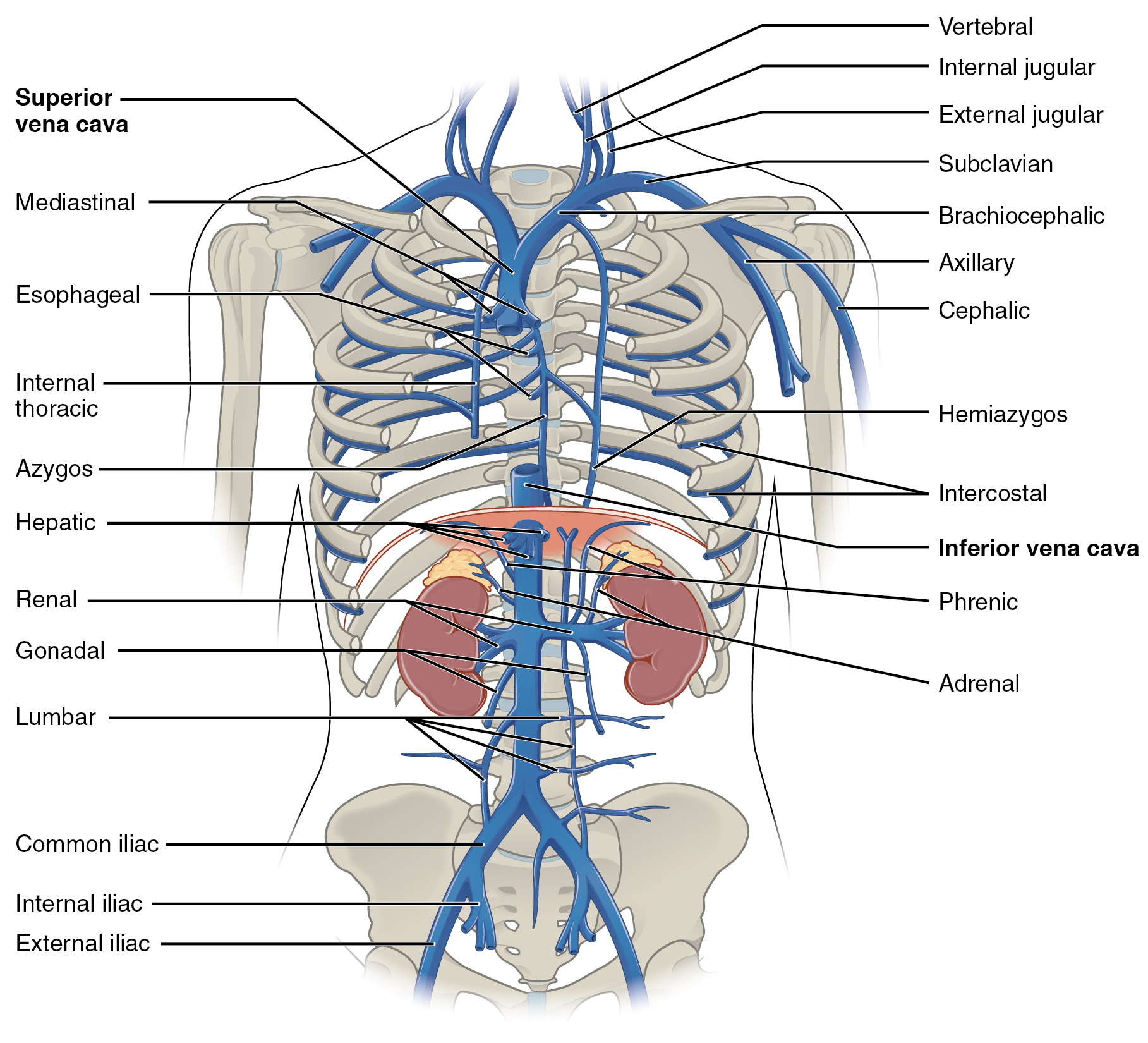
 Anatomy Of Major Abdominal Veins Inferior Vena Cava
/vascular-system-veins-56c87fa03df78cfb378b3e7c.jpg) What Is A Vein Definition Types And Illustration
What Is A Vein Definition Types And Illustration
 Visual Guide To Vein And Artery Problems
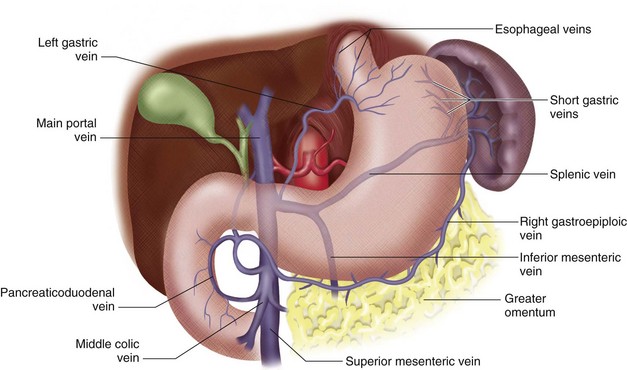
 Blood Supply To The Liver And Gallbladder
:max_bytes(150000):strip_icc()/arterial_system-59a5bdab68e1a200136f1b53.jpg) What Is A Vein Definition Types And Illustration
What Is A Vein Definition Types And Illustration
 Artery Vein And Capillary Anatomical Structure In Detail
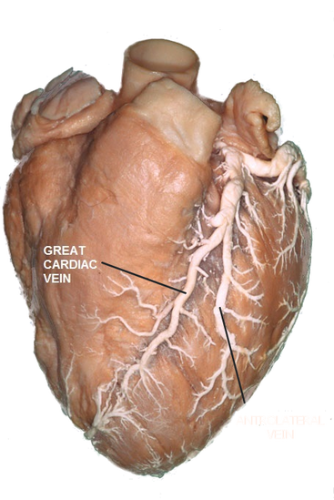 Cardiac Veins Cthsurgery Com
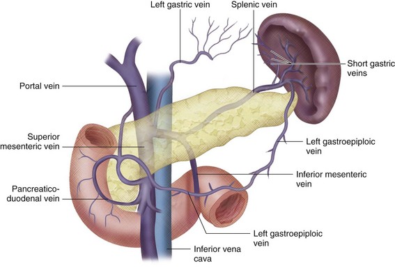
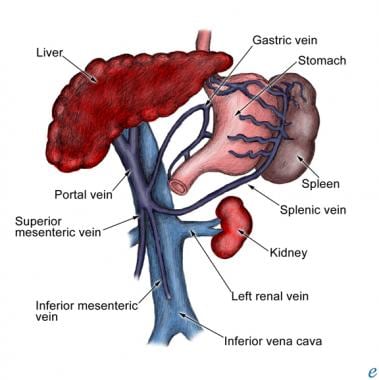 What Anatomy Is Relevant To Portal Hypertension
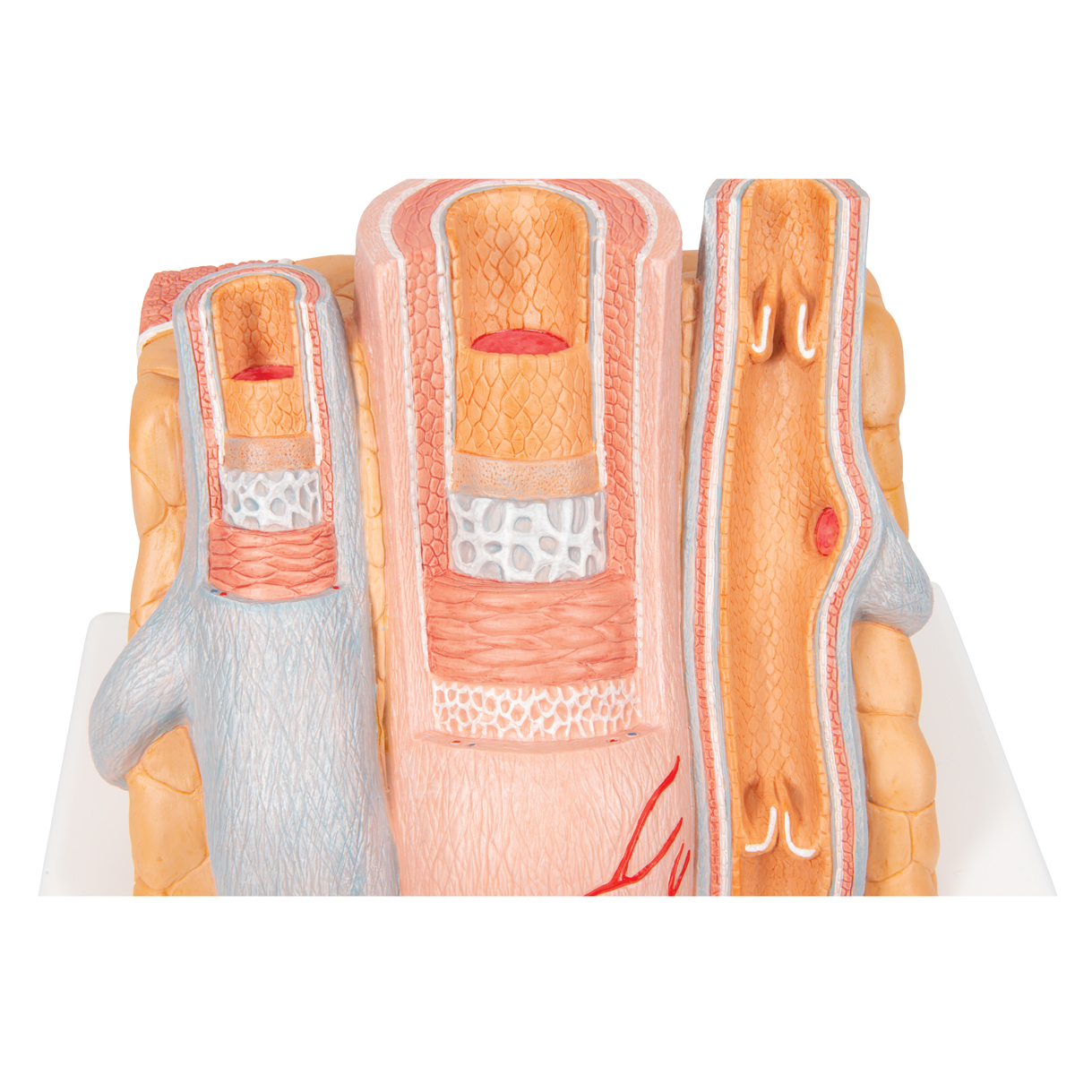 Anatomical Artery Vein Model Anatomy Of The Artery
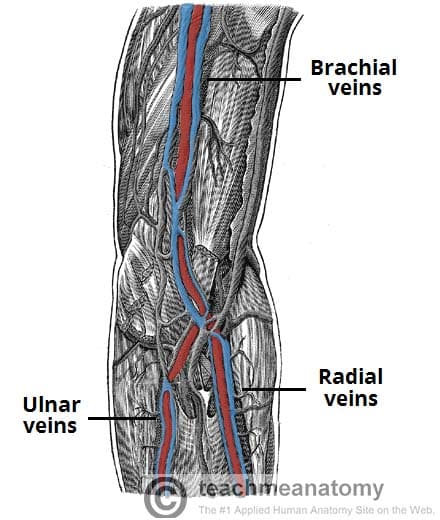 Venous Drainage Of The Upper Limb Basilic Cephalic
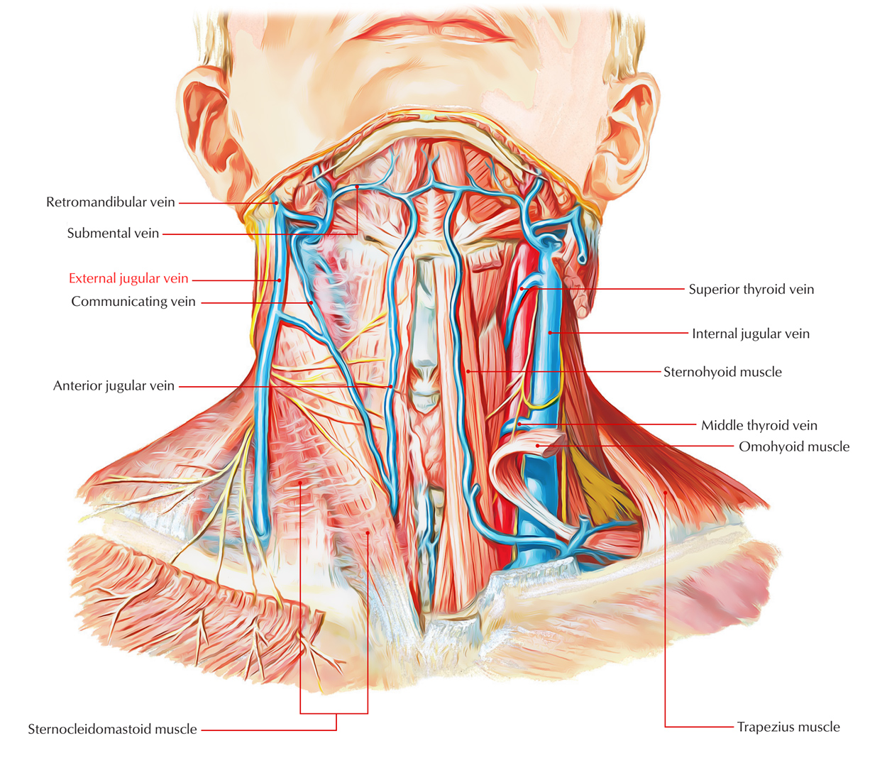
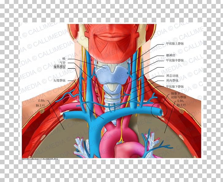
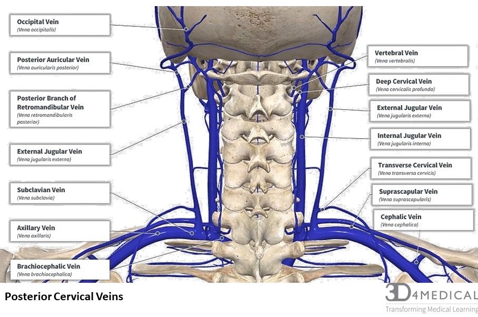
 The Veins Of The Neck Human Anatomy
 In The Human Umbilical Cord Are There 2 Arteries And 1 Vein
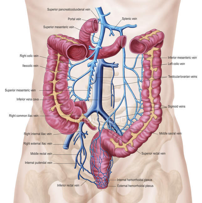
 Venous Anatomy Of The Abdomen And Pelvis Clinical Gate
 Venous Anatomy Sciencedirect
 Anatomy Of The Superior Mesenteric Vein Smv Portal Vein
 Veins In The Body Anatomy Human Body Veins Are Blood Vessels
 20 5 Circulatory Pathways Anatomy And Physiology
 Venous Anatomy Of The Abdomen And Pelvis Clinical Gate
 Anterior Triangle Of The Neck Posterior Triangle Of The Neck
![]() Veins Of The Brain Anatomy And Clinical Notes Kenhub
Veins Of The Brain Anatomy And Clinical Notes Kenhub
 Are The Jugular Vein And Carotid Artery Present On Both
![]() Veins Of The Brain Anatomy And Clinical Notes Kenhub
Veins Of The Brain Anatomy And Clinical Notes Kenhub
 Axillary Vein And Its Tributaries Upper Limb Gross Anatomy Dr G Bhanu Prakash Medical Animation
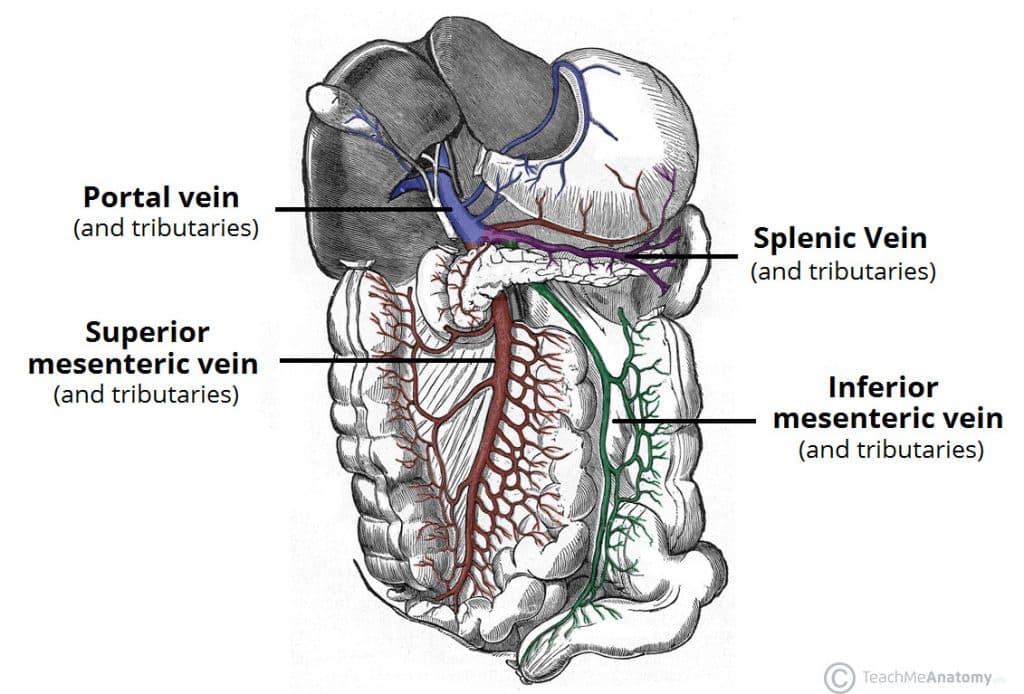 Venous Drainage Of The Abdomen Teachmeanatomy
 Superficial Vein An Overview Sciencedirect Topics
 Section 2 Anatomy And Physiology
 Not In The Same Vein I Spy Physiology Blog
 Nerves Blood Vessels And Lymph Advanced Anatomy 2nd Ed
 Anatomy Of Human Abdominal Vein System Blood Vessels
Belum ada Komentar untuk "Anatomy Of A Vein"
Posting Komentar