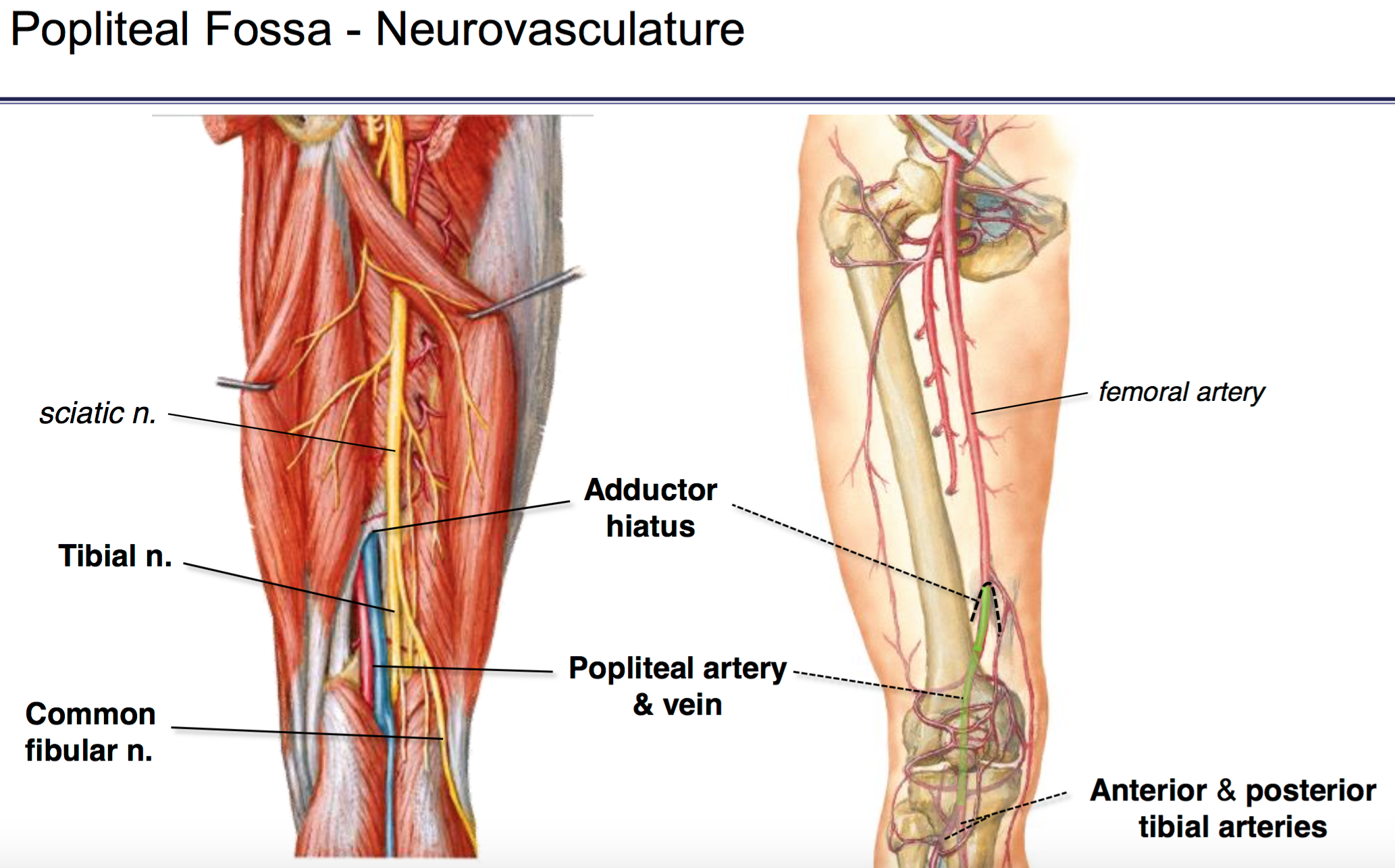
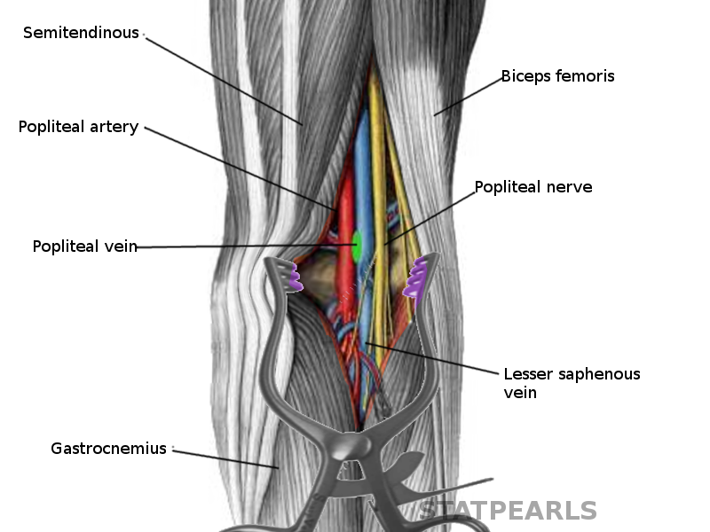
Popliteal Anatomy
Its courses near the adductor canal and the adductor hiatus distinctive open areas inside the thigh. Blood vessels are located deep to the nerves within the fossa and include.
 Anatomy Of The Popliteal Fossa Art As Applied To Medicine
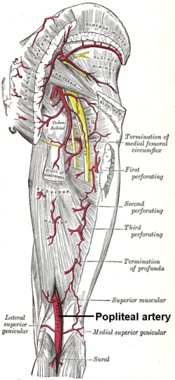
The popliteal artery is a deeply placed continuation of the femoral artery after it passes through alcocks canal or opening in the distal portion of the adductor magnus muscle.

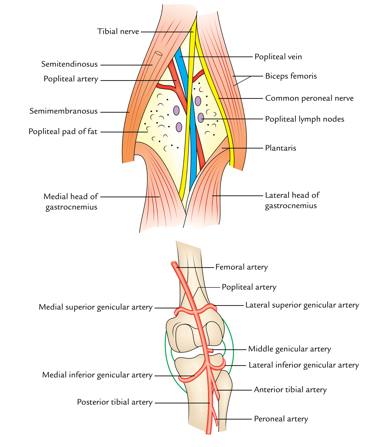
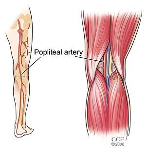
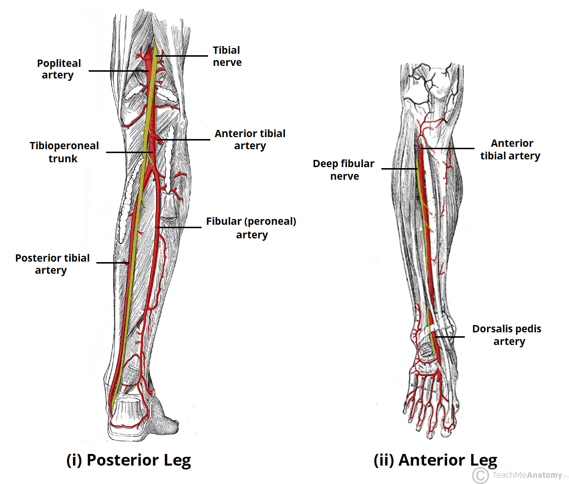
Popliteal anatomy. The popliteal artery located behind the knee is where the popliteal vein begins to extend. Its contents are medial to lateral. At its far end it splits into the anterior and posterior tibial arteries.
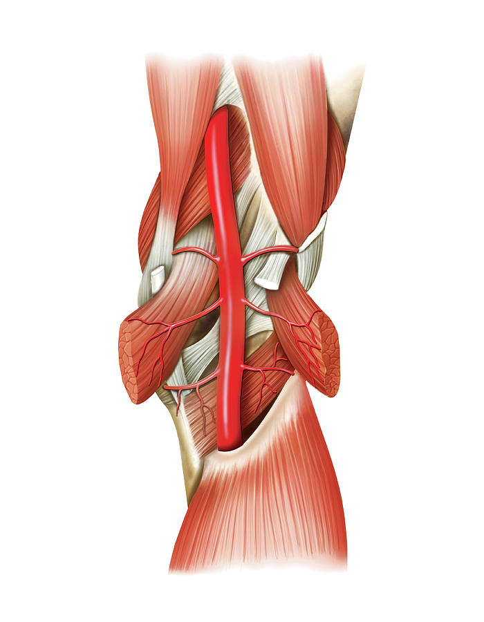
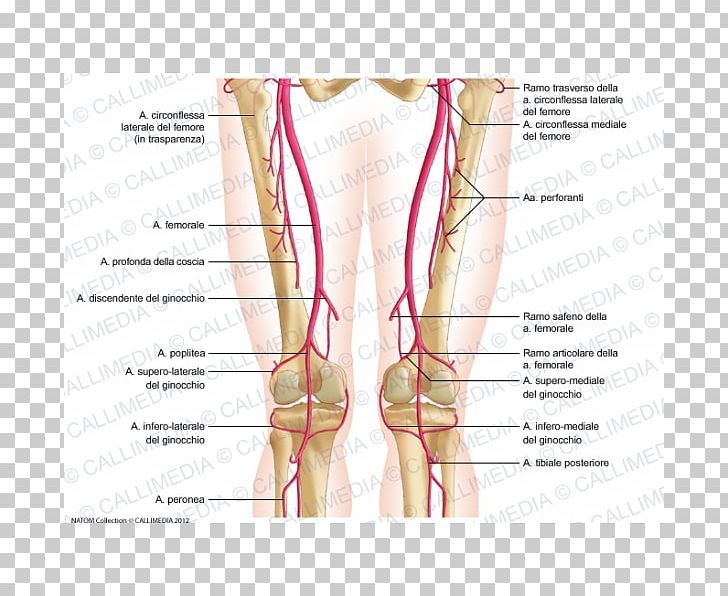
The popliteal artery is the continuation of the femoral artery that begins at the level of the adductor hiatus in the adductor magnus muscle of the thigh. The bones of the popliteal fossa are the femur and the tibia. It is located in the knee and the back of the leg.
This arterys primary job is to deliver blood to the bones and tendons of the knee. The popliteal vein drains the peroneal vein before it reaches the knee joint where it becomes the. The popliteal artery branches off from the femoral artery at the level.
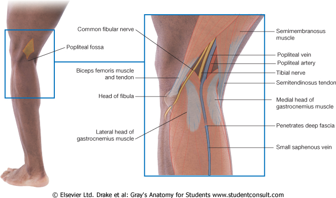
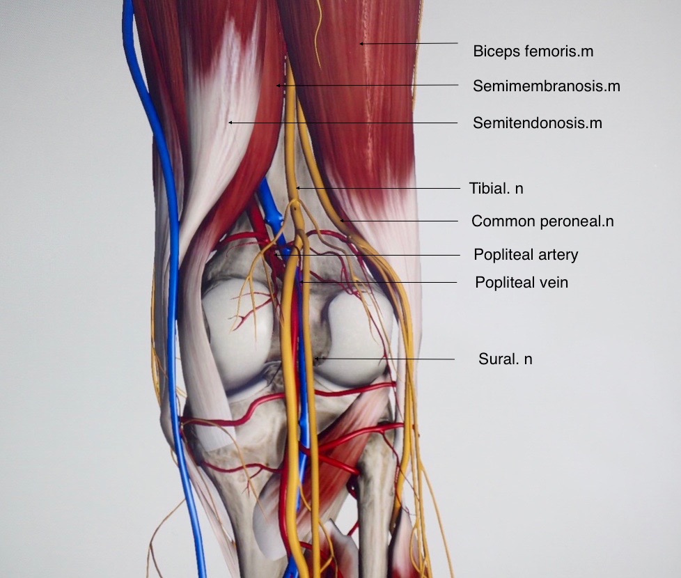
The popliteal fossa sometimes referred to as the kneepit or hough1 is a shallow depression located at the back of the knee joint. Common fibular nerve common peroneal nerve. During its course the popliteal artery branches into other significant blood vessels.
As it continues down it runs across the popliteal fossa posterior to the knee joint. The area of depression located at the back of the knee joint is called the popliteal fossa. Ebraheims educational animated video describes the anatomy of the back of the knee popliteal fossa.
The popliteal fossa is 25 cm wide and mainly consists of fat tissue. It lies medial to the popliteal artery in the lower part of the fossa posterior to the artery in the middle and posterolateral in the upper part of the fossa. Popliteal vein begins at the lower border of the popliteus by the union of veins accompanying the anterior and posterior tibial arteries.
The superomedial aspect of the popliteal fossa is bounded by the semimembranosus and. Several conditions are closely. A major artery that runs mid thigh to mid calf behind the knee anatomy.
It courses through the popliteal fossa and ends at the lower border of the popliteus muscle where it branches into the anterior and posterior tibial arteries.
 Functional Popliteal Artery Entrapment Syndrome A Review Of
 Pain Behind Knee Why It Hurts In Back Of Or Under Your Kneecap
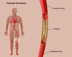 Femoral Popliteal Bypass Stanford Health Care
 Easy Notes On Popliteal Fossa Learn In Just 4 Minutes
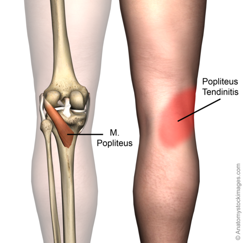 Popliteus Tendinopathy Physiopedia
 6 Anatomy Of Popliteal Fossa
 3d Printed Popliteal Fossa Model
 Knee Posterior Approach Approaches Orthobullets
 Popliteal Artery Anatomy And Course Bone And Spine
 Popliteal Artery Wikipedia
 The Popliteal Artery Human Anatomy
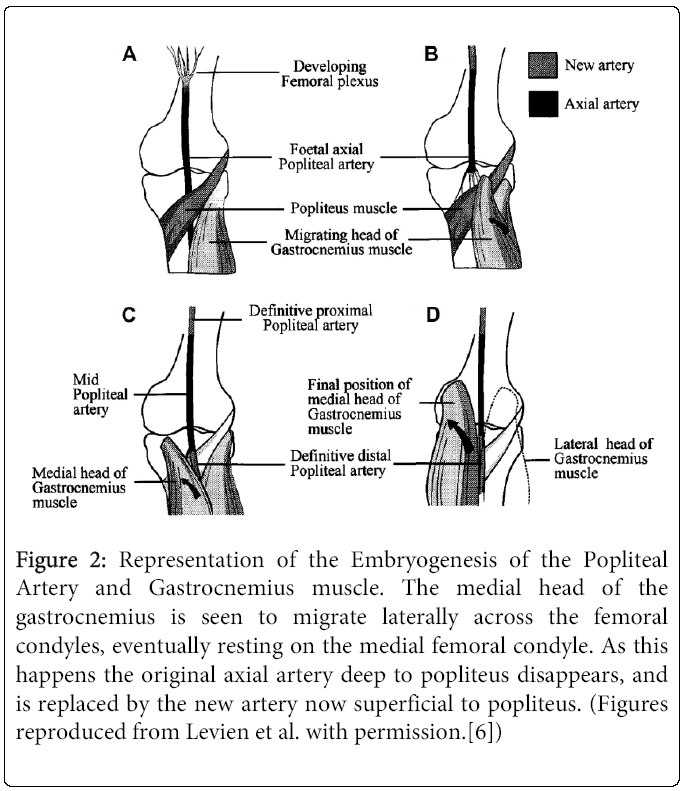
 The Evidence Based Surgical Anatomy Of The Popliteal Artery
 3d Printed Popliteal Fossa Distal Thigh And Proximal Leg
 Popliteal Artery
 Gastrocnemius Muscle An Overview Sciencedirect Topics
 6 Anatomy Of Popliteal Fossa
 Arteries Of The Lower Limb Thigh Leg Foot Teachmeanatomy
 Left Popliteal Vein The Anatomy Of The Veins Visual Guid
![]() Popliteal Artery Anatomy Branches Location And Course
Popliteal Artery Anatomy Branches Location And Course
 Human Leg Popliteal Artery Femoral Artery Knee Png Clipart
 Popliteal Anatomy Exhibits
 Popliteal Vein Wikipedia
 Anatomy Of The Right Popliteal Fossa The Dashed Line
 Untitled Document
 15074 01x Right Knee And Popliteal Artery Anatomy Exhibits
Belum ada Komentar untuk "Popliteal Anatomy"
Posting Komentar