Venous System Anatomy
This pressure ensures that veins carry blood to the heart and arteries transport it away. Veins are blood vessels that carry blood towards the heart.
 The Venous System Of The Male Human Body 1543 At Science
Most carry deoxygenated blood from the tissues back to the heart but the pulmonary and umbilical veins both carry oxygenated blood to the heart.

Venous system anatomy. The anatomy and physiology of venous return are described with an emphasis on the differences between standing and walking and the interplay between the venous systems of both the foot and the calf. The deep venous system includes the iliac femoral popliteal and deep femoral veins. Grossly the venous system is composed of venules and small and great veins which serve to return blood from tissues to the heart see the image below.
The cerebral venous system somewhat unlike the majority of the rest of the body does not even remotely follow the cerebral arterial system. Vein structure the walls of your veins are made up of three different layers. A short wide vein that carries blood to the liver from the organs of the digestive system.
The superficial subcutaneous venous system in the legs includes the long saphenous vein and the short saphenous vein. The reticular veins a network of veins parallel to the skin surface and lying between the saphenous fascia and dermis drain the lower extremity skin and subcutaneous tissue. The cortical veins lie superficially unlike cortical arteries and are adherent to the deep surface of the arachnoid mater so that they keep the sulci open 2.
The venous system refers to the network of veins that work to deliver deoxygenated blood back to your heart. Choose from 500 different sets of venous system anatomy flashcards on quizlet. It transports the blood from the surface skin and subcutaneous tissues where it collects in the deep veins.
The systemic venous system brings deoxygenated blood from tissues and organs back to the right atrium of the heart whereas the pulmonary venous system brings oxygenated blood from the pulmonary circulation back to the left atrium of the heart. The external iliac vein runs deep and is joined by the internal iliac vein which drains blood from the pelvis forming the common iliac vein. Learn venous system anatomy with free interactive flashcards.
The circulatory system works thanks to constant pressure from the heart and valves throughout the body. Anatomy of the venous system. The superficial venous system includes the reticular veins as well as the great greater and small lesser saphenous veins and their tributaries.
The left common iliac vein runs deep to the right common iliac artery to drain into the vena cava which lies to the right of the aorta.
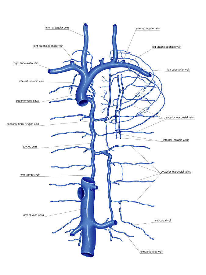 Venous System Of The Torso
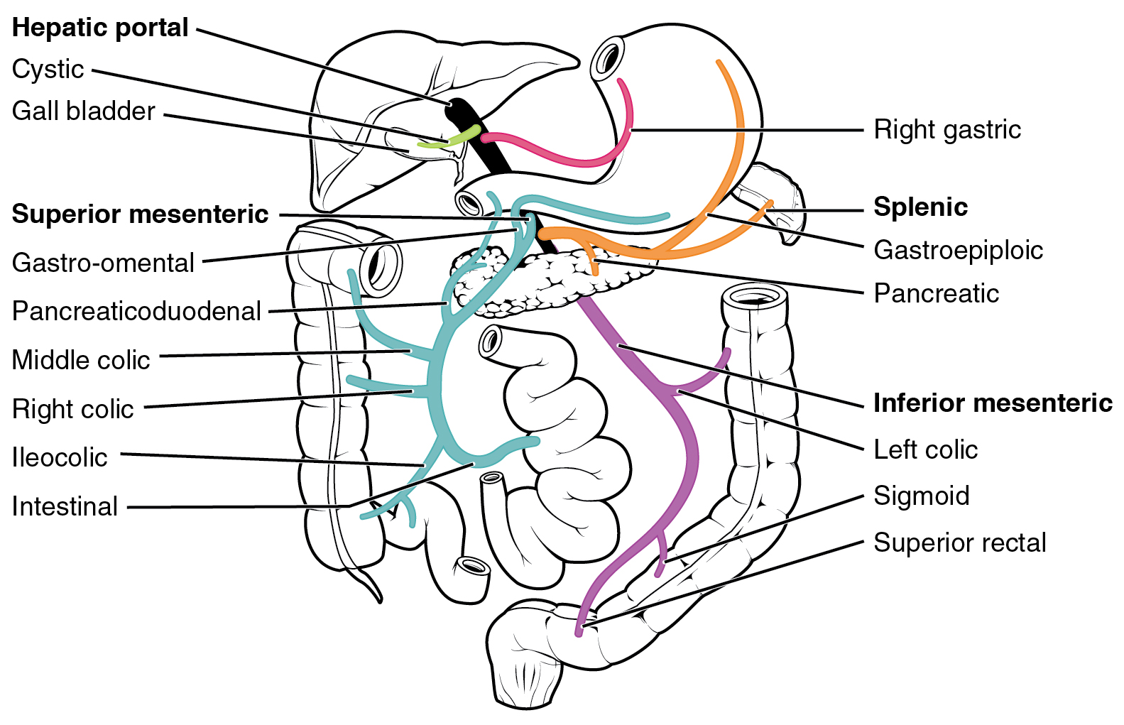
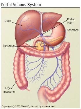
 Liver Cirrhosis Liver Cirrhosis The Anatomy Of The Portal
 Venous System
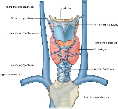
 Science Source Venous System Of The Head And Neck
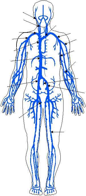
 20 Systemic Venous System Png Cliparts For Free Download


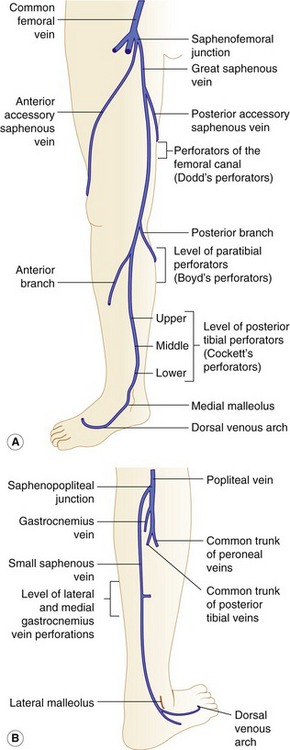 Anatomy Of The Lower Limb Venous System And Assessment Of
 20 5 Circulatory Pathways Anatomy And Physiology
 Venous System Of The Abdomen Art Print
 Basic Anatomy Of The Venous System Of The Lower Extremities
 Pediagenosis
 Venous System

 Venous System
 Azygos Venous System Anatomy Ct Pulmonary Angiography
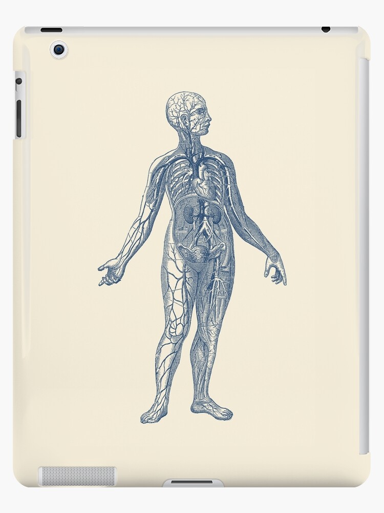 Venous System Diagram Vintage Anatomy Ipad Case Skin By Vaposters
 Vein Wikipedia
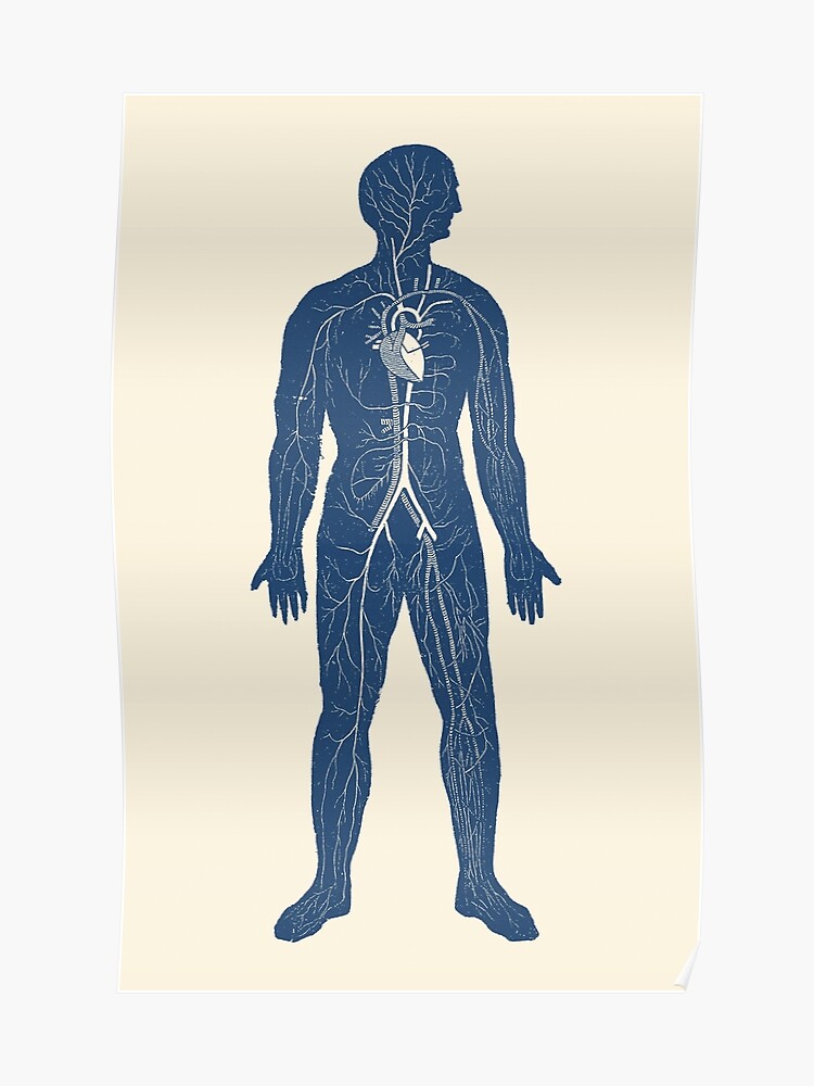 Venous Circulatory System Anatomy Diagram Poster
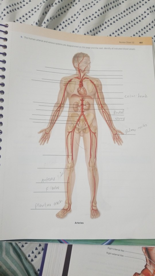 Solved 9 The Human Arterial And Venous Systems Are Diag
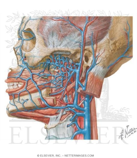 Vascular Supply Of The Face Venous Drainage

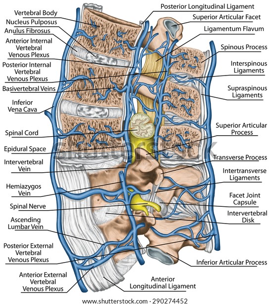 Internal External Vertebral Venous Plexuses Median Stock
Belum ada Komentar untuk "Venous System Anatomy"
Posting Komentar