Scotty Dog Anatomy
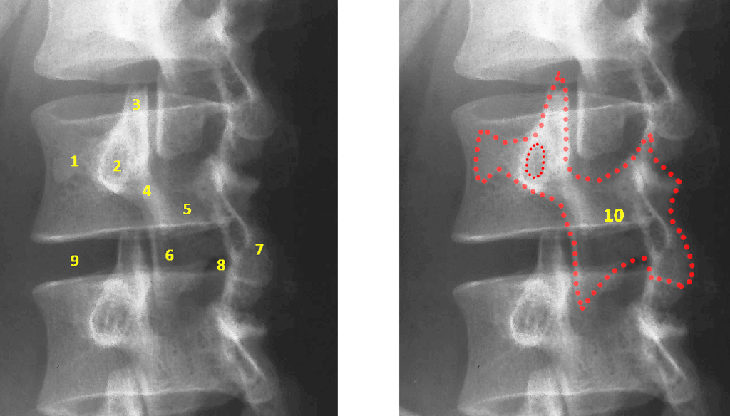
It is outlined above. On oblique views the posterior elements of the vertebra form the figure of a scottie dog with.
 Scotty Dog Sign Orthopaedicsone Articles Orthopaedicsone
Anatomy lessons this list provides links to other lessons that are found in the structure of the human body web site and which you may find useful in your studies.

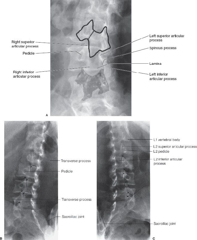
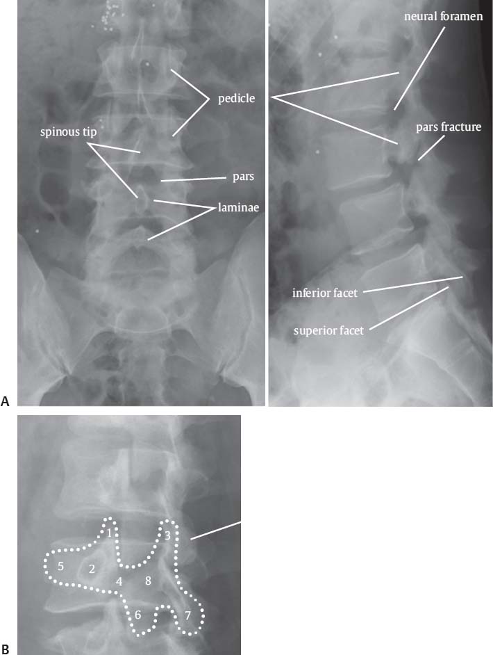
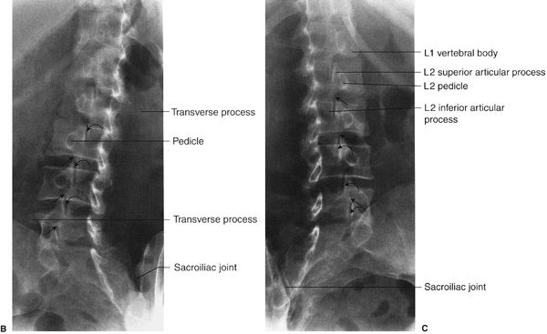
Scotty dog anatomy. The transverse process being the nose the pedicle forming the eye. Image filesize limited to low rez digital use normal lumbar spine oblique view sacroiliac joints spinous processes scottie dog pars interarticularis transverse process superior facet inferior facet joint lamina pedicle back plain x ray. The scottie dog sign refers to the normal appearance of the lumbar spine when seen on oblique radiographic projection.
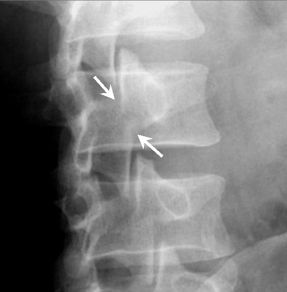
Radiology student radiology schools radiology humor radiologic technologist rad tech medical imaging anatomy images medical field dog anatomy. The pars fracture is seen as a collar around the dogs neck. When we look at the spine from the side we can imagine a scotty dog.
We will see this below in an x ray example. On oblique views the poste on oblique views the poste. His head is long in proportion to his size.
Scotty dog anatomy learn with flashcards games and more for free. He has a hard wiry weather resistant coat and a. Instability and movement can cause the neck to widen.
Part of the net profits from this shop go to scottish terrier emergency care and uk westie rehoming to help scottie and westies who are searching for a new forever home. Whether you love scottie and westie puppies or show dogs this is the place for you. Scotty dog x ray anatomysagitla lumbar spine x ray anatomy scotty dog.
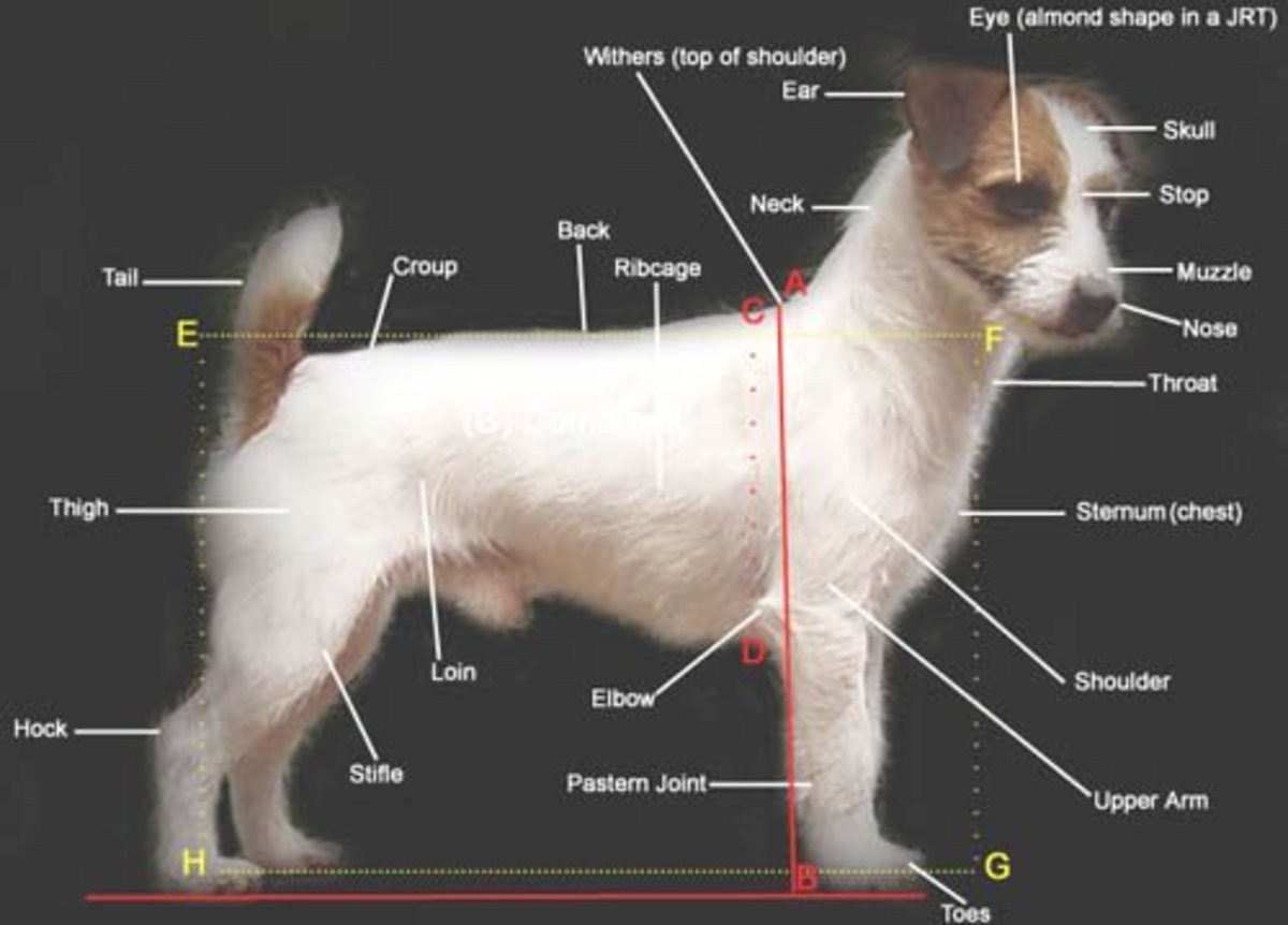
The scottish terrier is a small compact short legged sturdily built dog of good bone and substance. Like you we are dedicated to our dogs and know that you will like what we have to offer. Scottie dog sign spine the scottie dog sign refers to the normal appearance of the lumbar spine when seen on oblique radiographic projection.
Defect in the pars interarticularis disclaimers. The information in this video only represents the knowledge of the individuals depicted in it and not those of any institution or other. The lessons are arranged according to region.
 Radiological Anatomy Of The Spine
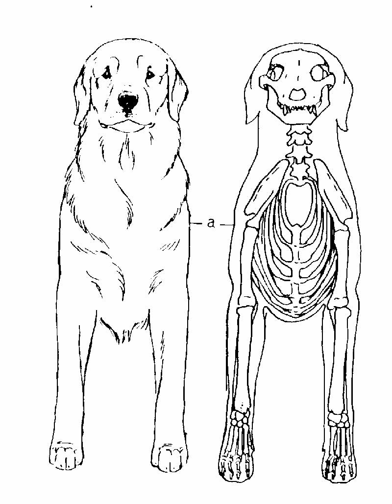
 Digestive System Of The Dog
 Neolithic Dog Recreated Using An Ancient Skull Big Think
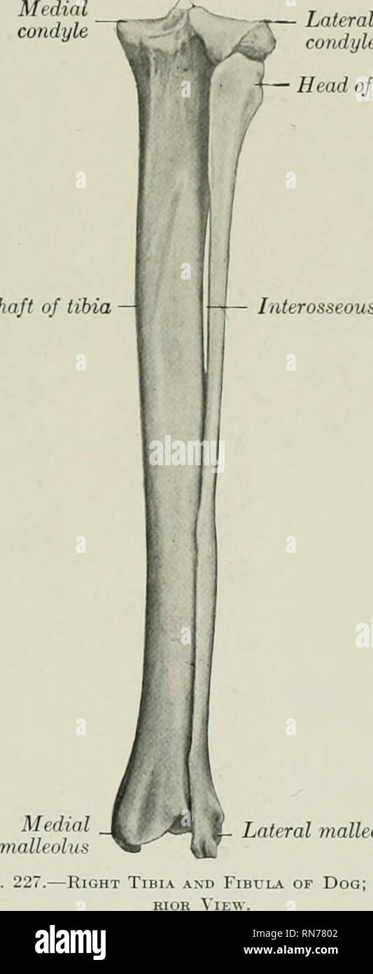
 Bone Radiologic Anatomy Bones And Joints
Z Joint Anatomy A Relevant Anatomy Nr Nerve Root Iap
 Bone Radiologic Anatomy Bones And Joints
 48 Common Health Problems Found In Jack Russell Terriers
 A Oblique Fluoroscopic View Scottie Dog Projection
 Radiological Anatomy Of The Spine
 Structure And Movement Pt 1 Breeding Better Dogs
 Scotty Dog Radiology Scotty Dog X Ray Anatomy Sagitla
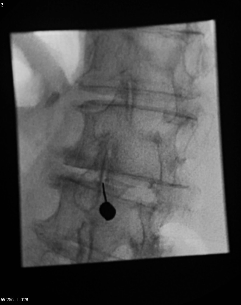 Facet Joint Injection Scotty Dog Radiology Case
 Learning Radiology Spondylolysis
 9 Spine And Pelvis Radiology Key
 Pars Interarticularis Anatomy And Significance Bone And Spine
 Lumbar Nerves An Overview Sciencedirect Topics
 Scotty Dog Sticker
 Scotty Dog Normal Anatomy Pars Interarticularis Ridley
 Lumbar Spine Annotated Oblique Projection Radiology Case
 1 Spinal Anatomy And Approaches Neupsy Key
 Vertebral Column Anterior Oblique View Scotty Dog
 Scottish Terrier Dog Breed Information Pictures
 Spine And Pelvis Radiology Key
 Docking Dog Wikipedia
 Little White Coats What Is The Clinical Significance Of A
 Classic Oblique View Optimized For The Scotty Dog Image
 Transforaminal Epidural Steroid Injection Atlas Of Pain
 Pars Defects Spondylolysis Spondylolisthesis Lumbar
 Resembles Dog Stock Photos Resembles Dog Stock Images Alamy
 Digestive System Of The Dog
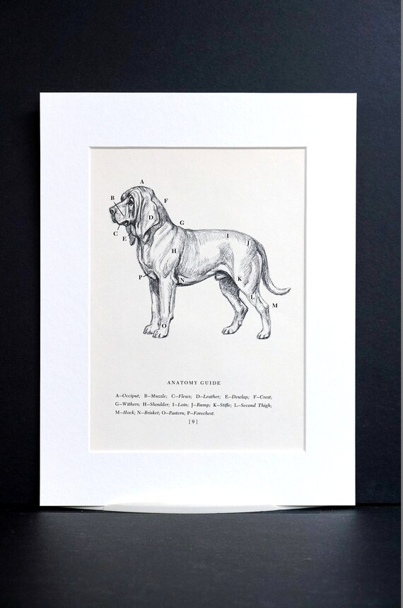 Bloodhound Dog Anatomy 1953 Edwin Megargee Veterinarian Veterinary Graduate Professionally Matted Vintage Dog Print Ready To Frame Wall Art
 Scotty Dog Youtube
 Back Problem Spondylolysis Pars Fracture Treatment
 Medpix Case Bilateral Pars Fracture Of L5 With Grade I


Belum ada Komentar untuk "Scotty Dog Anatomy"
Posting Komentar