Foot Anatomy Sole
Proximal interphalangeal joint pip the joint in the middle of the toe. Distal phalangeal joint dp the joint closest to the tip of the toe.
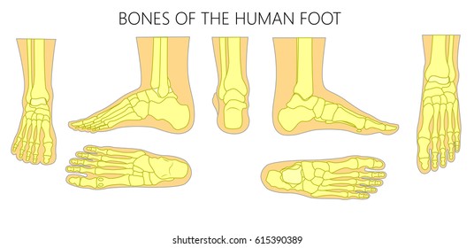 Foot Bones Photos 40 827 Foot Stock Image Results
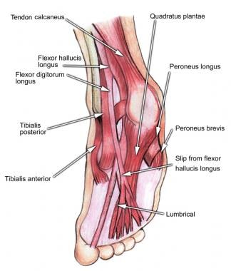
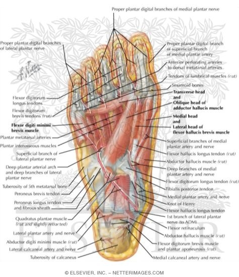
Central muscles of sole of the foot.
Foot anatomy sole. If studying by layers we can organise these muscles into four primary layers. The tendon of fibularis longus. Except for the big toe each of the toes has three joints which include.
There are 10 intrinsic muscles located in the sole of the foot. The foot is the lowermost point of the human leg. The foot contains 26 bones 33 joints and over 100 tendons muscles and ligaments.
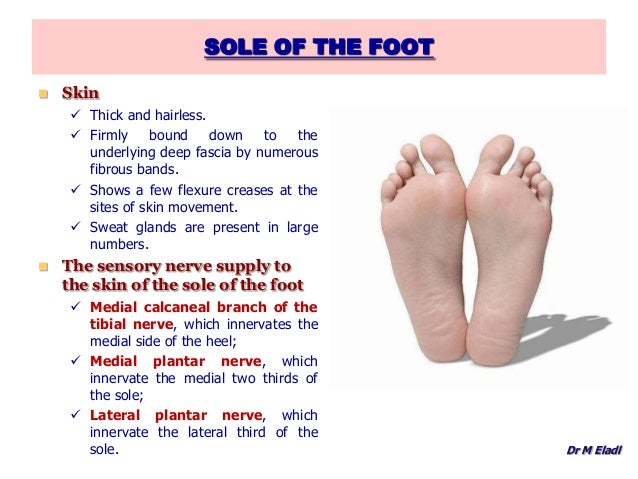
All the muscles are innervated either by the medial plantar nerve or the lateral plantar nerve which are both branches of the tibial nerve. Most superficial of all the layers. This may sound like overkill for a flat structure that supports your weight but you may not realize how much work your foot does.
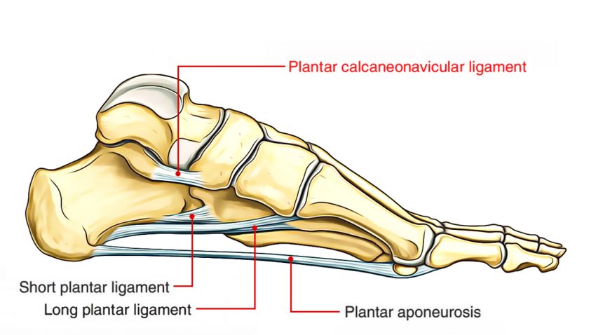
The muscles acting on the foot can be classified into extrinsic muscles those originating on the anterior or posterior aspect of the lower leg and intrinsic muscles originating on the dorsal top or plantar base aspects of the foot. The tendons of several extrinsic foot muscle reach the sole. They act collectively to stabilise the arches of the foot and individually to control movement of the digits.
The tendons of the deep foot flexors in the posterior compartment of the leg tibialis posterior flexor digitorum longus and flexor hallucis longus passes behind the medial malleolus into the sole. Metatarsophalangeal joint mcp the joint at the base of the toe. The plantar muscles of the foot are traditionally studied in either layers or groups.
Flexor digitorum brevis fdb abductor digiti minimi. Abductor hallucis flexor digitorum brevis abductor digiti minimi. The foots shape along with the bodys natural balance keeping systems make humans capable of not only walking but also running climbing and countless other activities.
Medically reviewed by healthline medical team on april 13 2015.
 Sole Foot Wikipedia
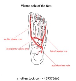 Vienna Sole Foot Twocolor Anatomical Illustrations Royalty
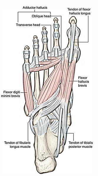 Easy Notes On Muscles Of The Foot Learn In Just 4 Minutes
 Diagram Of The Sole Of The Left Foot After Resection Of The
 Ledderhose Disease Physiopedia
 Plantar Foot Muscles Diagram Quizlet
 Muscles Of Sole Of Foot Third Layer
 Foot Sole Illustration Colored Vector Isolated On White
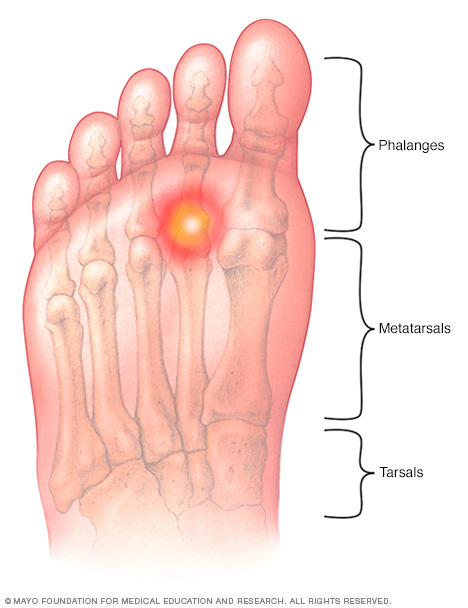 Metatarsalgia Disease Reference Guide Drugs Com
 Anatomy Of The Foot
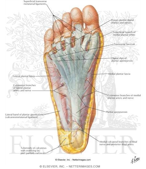 Sole Of Foot Superficial Dissection
Athletic Foot Injuries Background Epidemiology Functional
 Foot Anatomy Spokane Valley Wa Foot Doctor
 Anatomy 1 C4 L6 Back Of The Leg And Sole Of The Foot
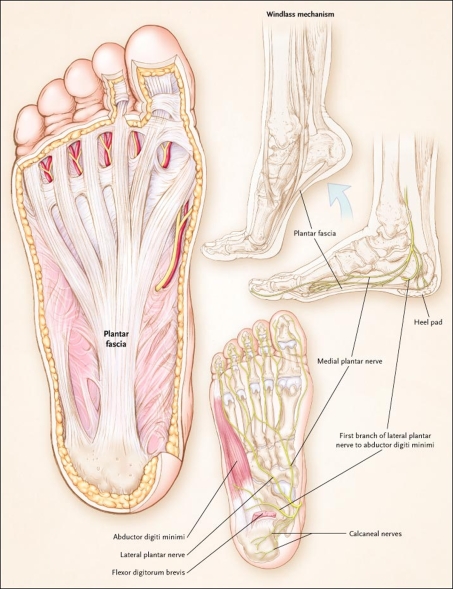
 Plantar Fasciitis Treatment Relief For Plantar Fasciitis
 Anatomy Of The Foot Pptx Anatomy Of The Foot Sub Title
 Human Foot Bone Model Foot Sole Joints Of Foot Ankle Tibia
 Ankle Foot Atlas Of Anatomy
 Anatomy Print Muscles Of The Sole Of The Foot First And
 Foot Pain Diagnosis Achilles Tendinitis Causes Home
 Foot And Ankle Patient Education
 Ecr 2007 C 638 Normal Sonographic Anatomy Of The Foot
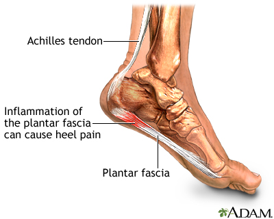 Plantar Fasciitis Medlineplus Medical Encyclopedia
 The Foot Advanced Anatomy 2nd Ed
 Sole Of Foot
 Ankle Foot Atlas Of Anatomy
Belum ada Komentar untuk "Foot Anatomy Sole"
Posting Komentar