Knee Anatomy Anterior
Knee anatomy anterior view and lateral view in detail in this image you will find articular cartilage patella anterior cruciate ligament acl lateral collateral ligament lcl quadriceps femur hamstring posterior cruciate ligament pcl meniscus medial collateral ligament mcl acl patellar ligament side view front view knee anatomy in it. Posteriorly the oblique popliteal ligament and arcuate popliteal ligament join the femur to the tibia and fibula of the lower leg.
 Ligaments Of The Knee Knee Sports Orthobullets
Start studying anatomy of anterior right knee.
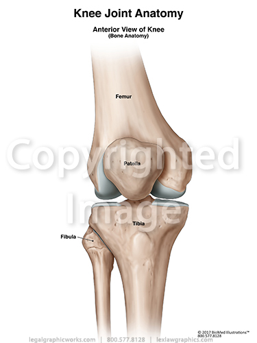
Knee anatomy anterior. The medial anatomy of the knee consists of several layers of structures that work together to provide stability and function. Anterior if facing the knee this is the front of the knee posterior if facing the knee this is the back of the knee. An acl tear often leads to the knee giving out and may require surgical repair.
Learn vocabulary terms and more with flashcards games and other study tools. The knee joint tibiofemoral joint is the largest and most complex joint of your body. The knee joint is a modified hinge joint because its primary movement is a uniaxial hinge movement that consists of three joints within a single synovial cavity.
Medial anatomy of the knee. Authors have used a variety of anatomic terms and descriptions that unfortunately have created ambiguity and confusion regarding this area of the knee. Pcl tears can cause pain swelling and knee instability.
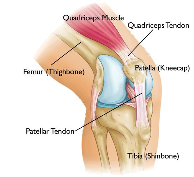
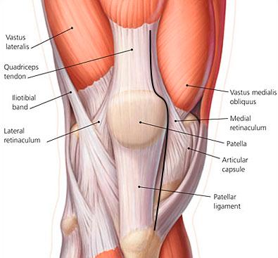
Anterior cruciate ligament anatomy. Medial anatomy of the knee. On the anterior surface of the knee the patella is held in place by the patellar ligament which extends from the inferior border of the patella to the tibial tuberosity of the tibia.
If used to describe the patella knee cap then it would refer to the side of the patella closest to the femur. The medial anatomy of the knee consists of several layers of structures that work together to provide stability and function. The knee joint is made up of three bonesthe femur thigh bone tibia shin bone and patella kneecap.
Acl anterior cruciate ligament strain or tear. Anterior knee painwhich simply means pain in the front of the kneeis two to seven times more common in women than men. To test for this you can perform an anterior drawer test where you attempt to pull the tibia forwards if it moves the ligament has been torn.
Pcl posterior cruciate ligament strain or tear. And it can have many causes. First a bit of anatomy.
The acl is responsible for a large part of the knees stability. Authors have used a variety of anatomic terms and descriptions that unfortunately have created ambiguity and confusion regarding this area of the knee. The anterior cruciate ligament acl can be torn by hyperextension of the knee joint or by the application of a large force to the back of the knee with the joint partly flexed.
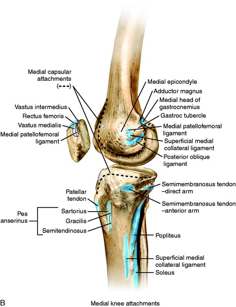 Medial And Anterior Knee Anatomy Musculoskeletal Key
 How To Keep Your Knees Safe And Injury Free During A Yoga
Anatomy Of The Knee Knee Specialist Fairfield Shelton
 Acl Solutions Acl Knee Anatomy And Diagram Images
 Knee Pain On The Inside Of Your Joint Causes Solutions
 Anterior Cruciate Ligament Acl Injury Orthopedics
 The Knee Anatomy Injuries Treatment And Rehabilitation
 Patella An Overview Sciencedirect Topics
 Anatomy Of Human Knee Joint Art Print
 Anatomy Of The Knee Bones Muscles Arteries Veins Nerves
 Matthew Boyle Orthopaedic Surgeon Knee Anatomy Knee
 Anterior View Of Knee Joint Bones
 Knee Joint Part 2 3d Anatomy Tutorial
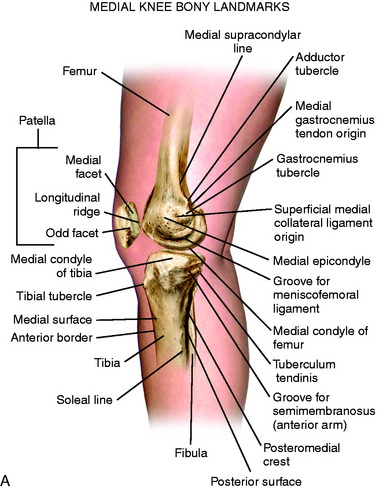 Medial And Anterior Knee Anatomy Musculoskeletal Key
 Anatomy Of Human Knee Joint Greeting Card
Anterior Knee Pain Essex Orthopaedics Sports Medicine
 Acl Tears Pinnacle Orthopaedics
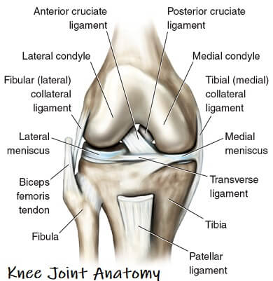 Knee Joint Anatomy Motion Knee Pain Explained
 Knee Joint Picture Image On Medicinenet Com
 Joints Ligaments And Connective Tissues Advanced Anatomy
 Adolescent Anterior Knee Pain Orthoinfo Aaos
 Tka Approaches Recon Orthobullets
 Anatomy Of The Knee Comprehensive Orthopaedics
Common Knee Injuries Orthoinfo Aaos
Belum ada Komentar untuk "Knee Anatomy Anterior"
Posting Komentar