Lower Extremity Vein Anatomy
The superficial veins are located within the subcutaneous tissue whilst the deep veins are found deep to the deep fascia. Additionally ultrasound is used to guide endovenous procedures such as foam sclerosant injection endovenous laser therapy evlt or radiofrequency ablation rfa for the treatment of superficial venous disease.
 Lower Extremity Vein Anatomy Venous Diseases Dallas Vein
Veins of the lower limb.

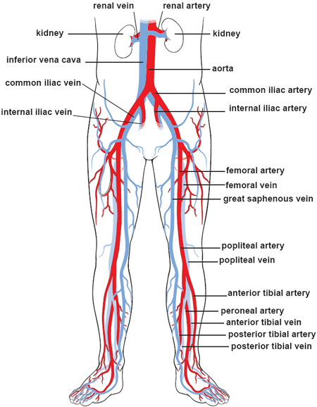
Lower extremity vein anatomy. The venous system of the lower extremities includes the deep veins which lie beneath the muscular fascia and drain the lower extremity muscles. The superficial veins which are above the deep fascia and drain the cutaneous microcirculation. Lower limb venous duplex imaging can be used for the assessment of patients with primary or recurrent varicose veins or for the investigation of patients with skin changes and venous ulceration.
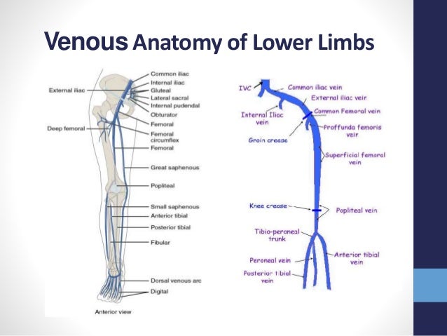
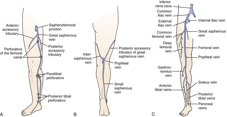
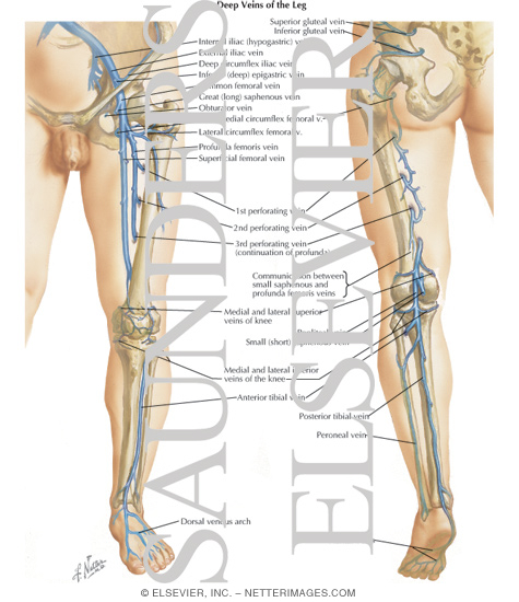
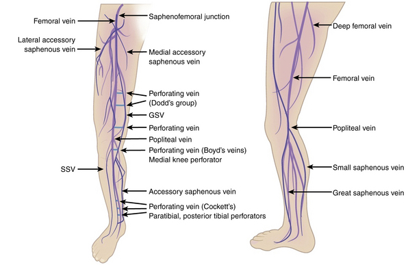
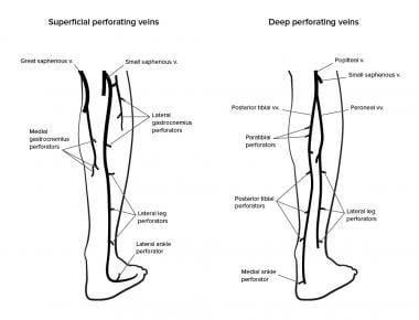
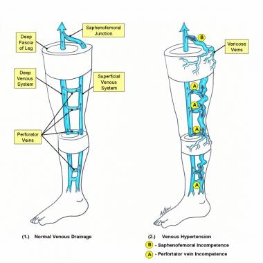
The lower extremity venous anatomy is divided into three interconnected systems. And the perforating veins that penetrate the muscular fascia and connect the superficial and deep veins. Deep superficial and perforating veins.
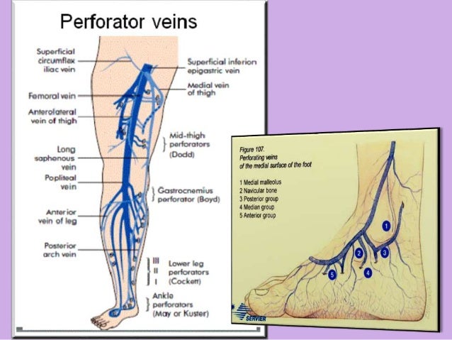
The veins rely on a more passive and indirect mechanism to help aid in the return of blood to the heart. Inguinal perforators drain into the femoral vein in the proximal thigh. The lower limb consists of two main types of veins.
Valves in superficial veins of the lower extremity usually are located near to the termination of major tributaries. Some valves are well developed with marked sinusoid dilation at their base others are more delicate in their structure. In us examination of the lower extremity veins knowledge of the venous anatomy as well as appropriate patient positioning and transducer placement are important for optimal imaging and accurate diagnosis.
In the pelvis and lower extremities blood in the veins has to fight gravity in order to flow in the correct direction. Lower extremity venous anatomy. The venous system of the lower extremities is divided into three groups.
The deep veins accompany the major arteries and their branches and are usually paired.
 Lower Extremity Venous Anatomy Google Search Medical
 Varicose Veins By M Fathy Zaidan
 3 Anatomy Of The Lower Extremity Veins Accessory Veins Are
 The Hemodynamics And Diagnosis Of Venous Disease Sciencedirect
 Basic Anatomy Of The Venous System Of The Lower Extremities
 Assessment And Management Of Older People With Venous Leg Ulcers
 Rosen Vein Lecture 102 Lower Extremity Vein Anatomy Vein
 Venous Physiology Clinical Gate
 Rosen Vein Lecture 102 Lower Extremity Vein Anatomy Vein
 Ultrasound Evaluation Of The Peripheral Vascular System
 Venous Drainage Of Lower Limb Ppt
 Veins Of The Lower Limb An Overview Sciencedirect Topics
 Pdf Ultrasonographic Anatomy Of The Lower Extremity
 Emergency Ultrasound

 Anatomy Of The Lower Extremity Veins Varicose Veins
 Diagram Showing The Venous Anatomy Of The Leg
 Venous Disorders Of The Lower Extremity

 Lower Limb Venous Anatomy Thoracic Key
 Varicose Veins Clinical Gate
 Lower Extremity Veins Techniques And Interpretation With How To Demonstration

 Venous Insufficiency Background Anatomy Pathophysiology
 Vessel Anatomy Veins Of The Lower Extremities Diagram
 Ultrasonography
 Femoral Vein Wikipedia
 Varicose Vein Surgery Practice Essentials Anatomy
Belum ada Komentar untuk "Lower Extremity Vein Anatomy"
Posting Komentar