Stifle Anatomy
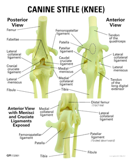
The largest joint in the body the knee moves like a hinge allowing you to sit squat walk or jump. The ligament prevents the tibia from rotating medially at the stifle varus deviation of the stifle when the limb is extended.
 Modified Technique For Medial Patellar Desmotomy In Donkey
It is held in place by a ligament at the bottom and a tendon on top.
Stifle anatomy. The stifle joint often simply stifle is a complex joint in the hind limbs of quadruped mammals such as the sheep horse or dog. This article also tells you how a normal knee works and provides resources for problems of the knee joint or its parts including knee injuries. Ligaments of the knee.
They act like strong ropes to connect bones. Cartilage of the knee. Patella the thick triangular bone that sits over the other bones at the front of the knee or kneecap.
The knee joint is the largest and one of the most complex joints in the human body. The lateral collateral ligament is taut when the stifle is extended and lax when the stifle is flexed. There are various muscles that control movement ligaments that give stability special cartilage to absorb pressure and various other structures to ensure smooth pain free movement.
Ligaments are tough and fibrous tissues. The knee is a modified hinge joint which permits flexion and extension as well as slight internal and external rotation. There are three bones that come together at the knee joint.
The acl is responsible for a large part of the knees stability. The knee consists of three bones. Anatomy of the knee bones around the knee.
These tough bands of soft tissue. Those connect to the femur and tibia. Knee anatomy is about the structure of the knee that is the parts that makeup the knee.
It is often termed a compound joint having tibiofemoral and patellofemoral components. The kneecap slides along a groove in the femur as the knee bends. Knee joint anatomy involves looking at each of the different structures in and around the knee.
An acl tear often leads to the knee giving out and may require surgical. Knee joint anatomy function and problems. Ligaments are structures that connect two bones together.
The knee is the joint where the bones of the lower and upper legs meet. These are crescent shaped discs that act as a cushion. May 21 2018 by 24 comments.
There are two types of cartilage of the knee joint. The femur thigh bone tibia shin bone and patella. It is the equivalent of the human knee and is often the largest synovial joint in the animals body.
The knee is vulnerable to injury and to the development of osteoarthritis. Acl anterior cruciate ligament strain or tear.
 Behind The Bit The Stifle The Mother Of All Joints
 Horse Anatomy Mobility Health
 Addressing Hock And Stifle Issues American Farriers Journal
 How To Perform Arthrocentesis Of The Compartments Of The
 Equine Stay Apparatus Hindlimb 3d Veterinary Anatomy Learning Ivala
 The Stifle Is Comparable To The Human Knee What My Mare
 Stifle Joint Stock Photos Stifle Joint Stock Images Alamy
 Pdf Magnetic Resonance Imaging Of The Equine Stifle
 Other Joint Disorders In Dogs Dog Owners Merck
 Canine Arthrology Illustrations
 Cruciate Ligament Rupture And Associated Injuries
 Canine Knee Model 9050 For Sale Anatomy Now
 Module 2 Exploring The Energetic Week 10 Whole Horse
 Patella And Sesamoid Bones Of The Stifle Ovam
 Anatomy Of The Canine Knee Stifle Joint Pet Guardian
 File The Anatomy Of The Horse A Dissection Guide 1922
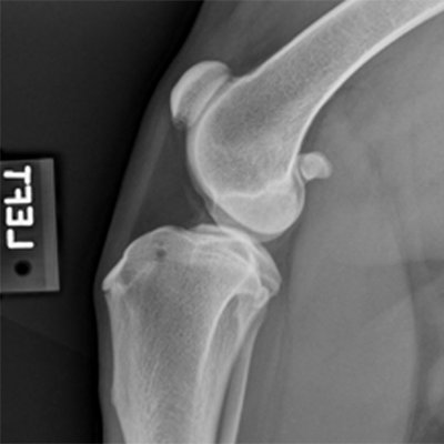
 Stifle Joint Anatomy Of The Dog On Ct
 Equine Stifle An Overview Sciencedirect Topics
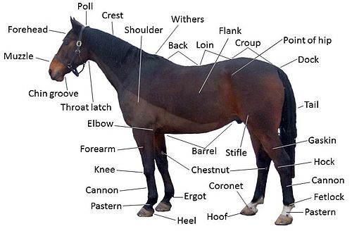 Equine Anatomy Wikipedia
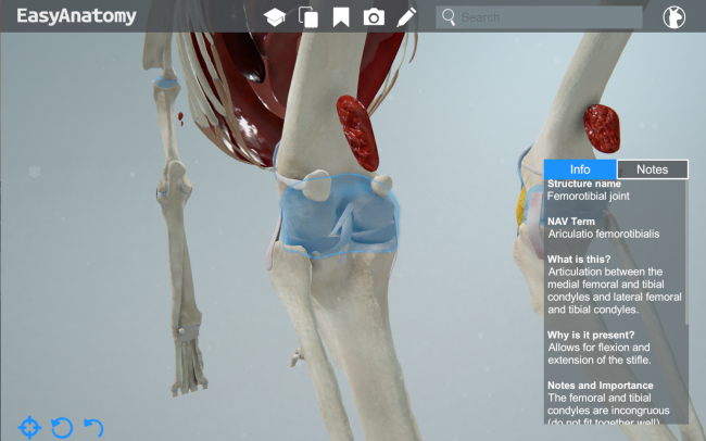 Anatomy Of The Canine Knee Easyanatomy
 Diagnosing Cranial Cruciate Ligament Pathology
 Joints Anatomy Physiology Wikivet English
 Diagnosing Cranial Cruciate Ligament Pathology
 Imaging Anatomy
 Canine Knee Model
 The Canine Stifle Sciencedirect


Belum ada Komentar untuk "Stifle Anatomy"
Posting Komentar