The Chest Anatomy
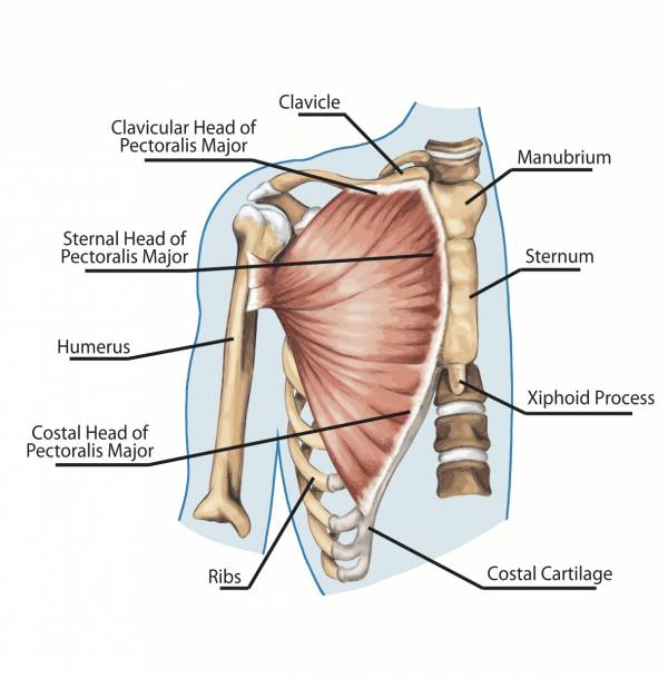
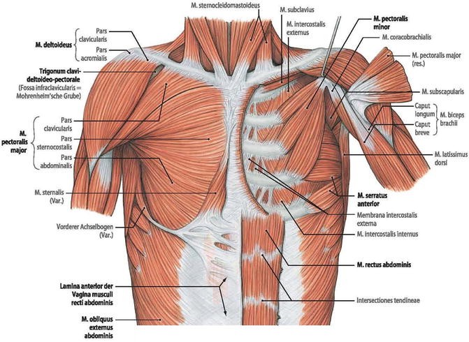
The thorax includes the thoracic cavity and the thoracic wall. This large fan shaped muscle stretches from the armpit up to the collarbone and down across the lower chest region on both sides of the chest.
 File Chest Anatomy Jpg Wikimedia Commons
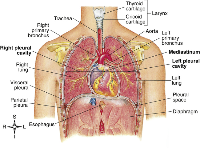
The thorax or chest is a part of the anatomy of humans and various other animals located between the neck and the abdomen.

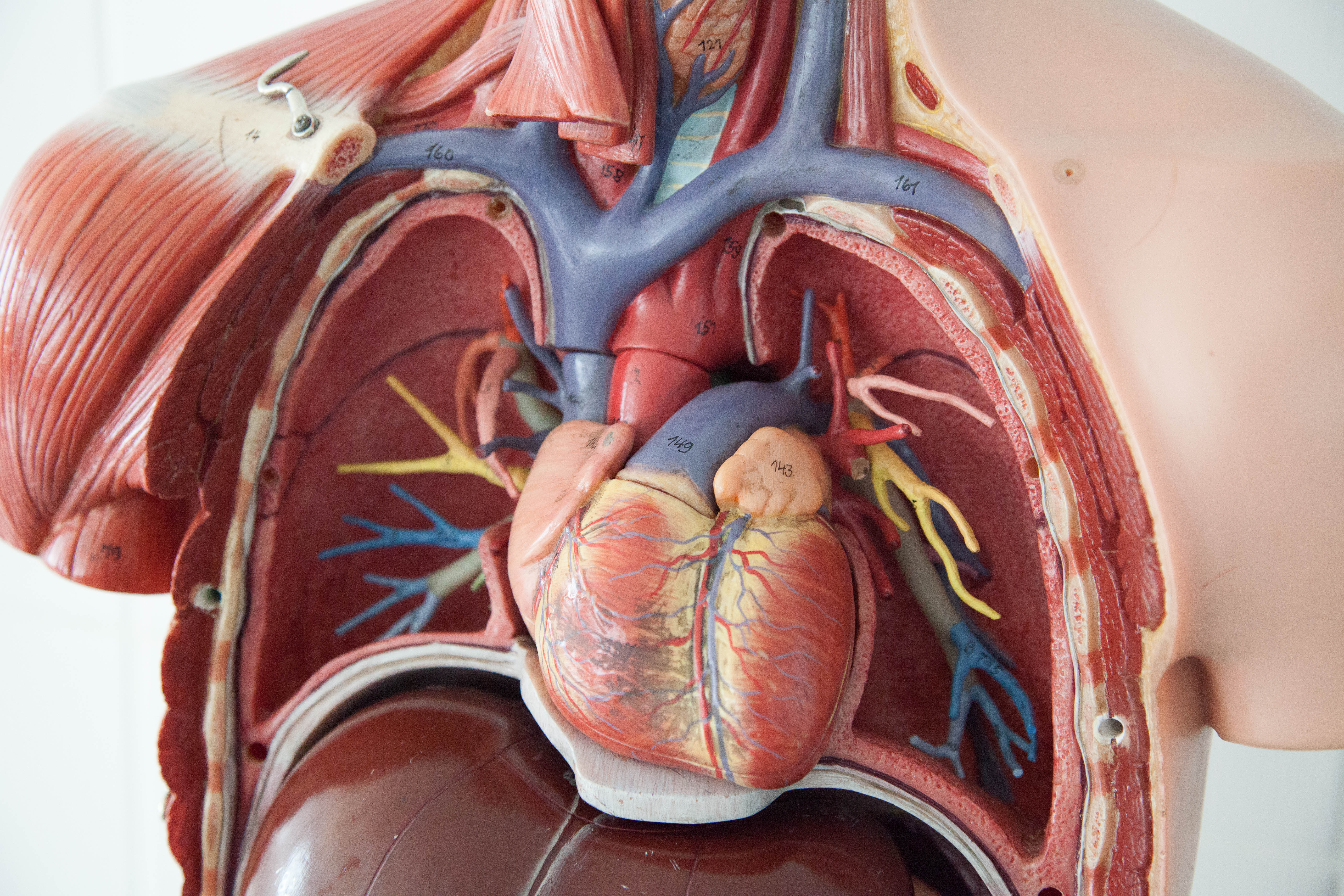
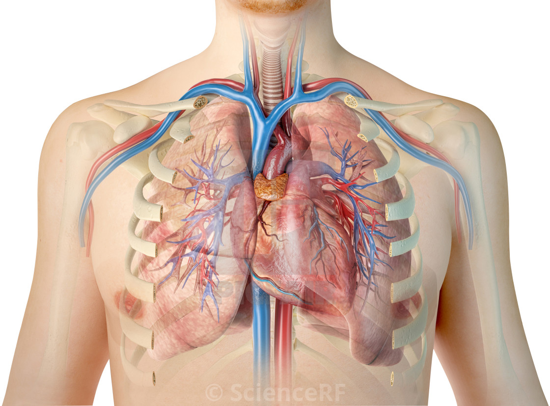
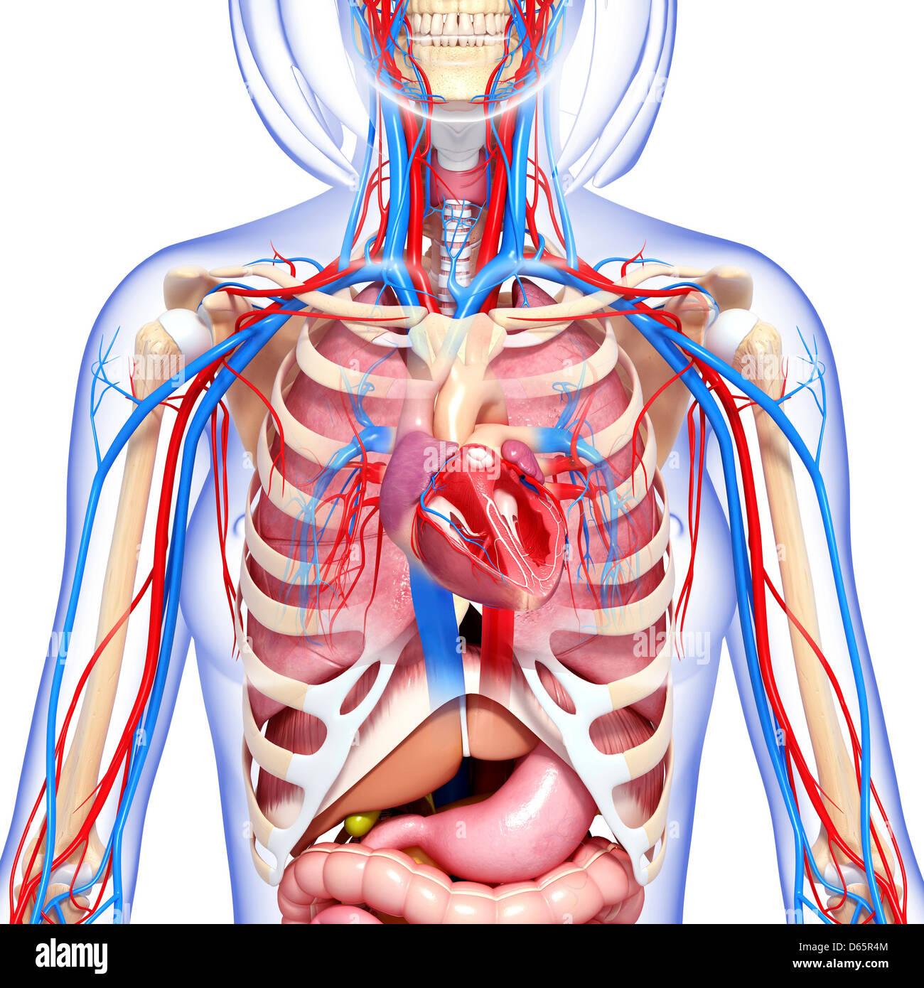
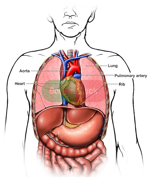
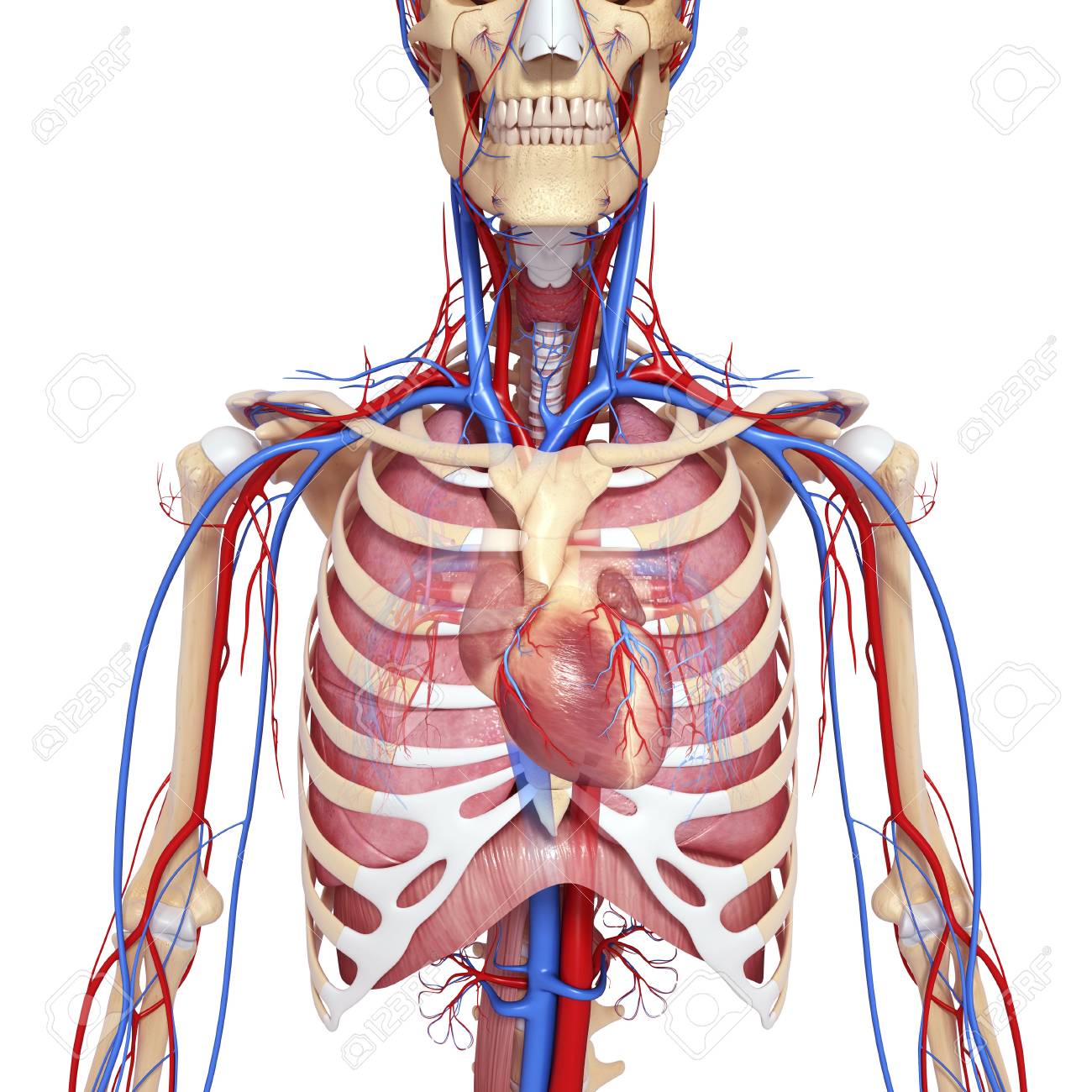
The chest anatomy. The circulatory system does most of its work inside the chest. The chest is the area of origin for many of the bodys systems as it houses organs such as the heart esophagus trachea lungs and thoracic diaphragm. It contains organs including the heart lungs and thymus gland as well as muscles and various other internal structures.
This synergy is a reference to dark souls in particular a message left by a player alluding to a characters large chest. Anatomy of the chest and the lungs. The pectoralis major is a large substantial fan shaped muscle.
Anatomy of a transverse ct of the thorax duration. Prior to the a farewell to arms update book of chest anatomy was a item. This mri chest thorax axial cross sectional anatomy tool is absolutely free to use.
Use the mouse scroll wheel to move the images up and down alternatively use the tiny arrows on both side of the image to move the images. The chest is part of a larger group of pushing muscles found in. Eva sweeney 84676 views.
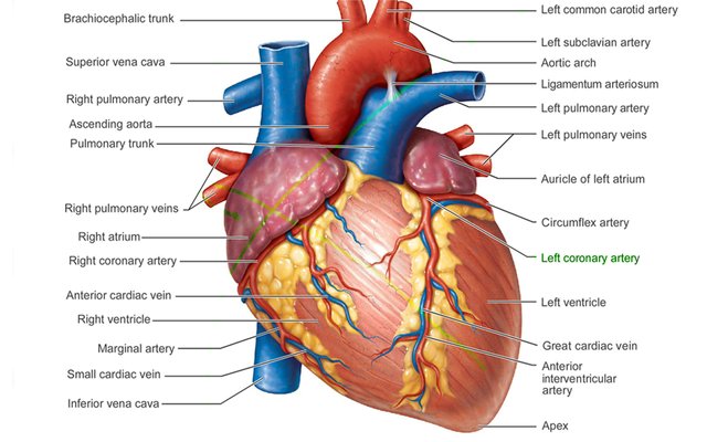
There the heart beats an average of 72 times a minute and circulates up to 2000 gallons of blood a day. The pectoralis minor is a thin triangular muscle that. The major muscle in the chest is the pectoralis major.
Anatomical illustrations this e anatomy module presents an illustrated anatomy of the lungs trachea bronchi pleural cavity and pulmonary vessels. The dermis is the under layer that contains sweat glands hair follicles blood vessels and more. All about the chest muscles function of the chest muscles.
This thoracic and pulmonary anatomy tool is especially designed for students of anatomy medical and paramedical studies. Chest the epidermis is the outermost layer that provides a protective waterproof seal over the body.
 Clinical Examination Of The Chest Wall
 Anatomy Of The Chest Medical Illustration Human Anatomy
 Human Chest Anatomy Illustration License Download Or
 Normal Female Anatomy Of The Chest Thoracic Cavity And
 Female Chest Anatomy Stock Photos Female Chest Anatomy
 Chest Anatomy What Are The Muscles And What Do They Do
 Thorax Wikipedia
 Location Of The Heart In The Chest Cavity Steemit
 Chest Muscles Anatomy Learn For Better Workouts
 Muscles Of The Chest And Upper Back
 Chest Anatomy Stock Photos Chest Anatomy Stock Images Alamy
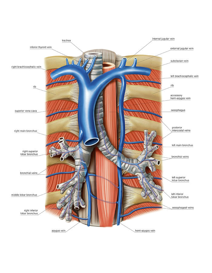 Venous System Of The Chest
![]() Anatomy Of The Back Spine And Back Muscles Kenhub
Anatomy Of The Back Spine And Back Muscles Kenhub
 Thorax Surface Anatomy 4 Edition

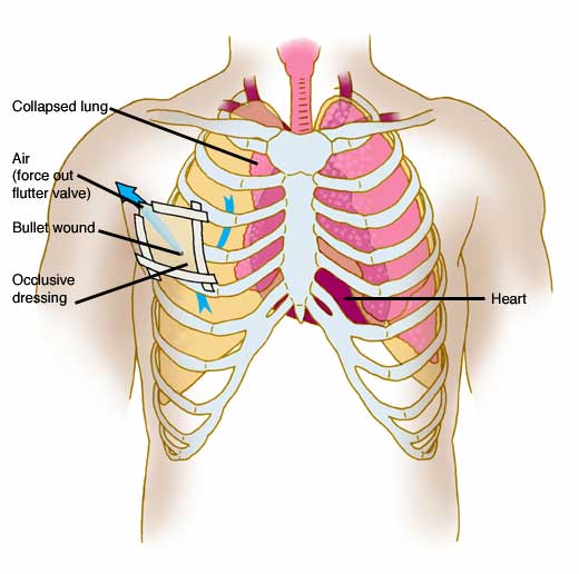 Anatomy Atlases Anatomy Of First Aid A Case Study Approach
 The Muscles Of The Chest And Upper Back Anatomy Medicine Com
 Anatomy Of The Thoracic Chest Organs Doctor Stock
 Figure 6 From The Anatomy Of The Ribs And The Sternum And
 Pin By Jr On Paramedic Emt Human Anatomy Physiology

Surgical Anatomy Of The Chest Wall Thoracic Key
 Solved Identify The Indicated Muscles Of The Chest And Ab
 Thorax Surface Anatomy 4 Edition
 Bony Surface Landmarks On The Anterior Chest Note The
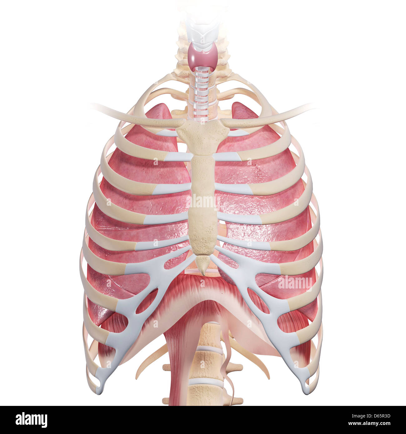
 Chest Anatomy Artwork
 Surgical Anatomy Of The Chest Wall Springerlink
 Anatomy Of The Breast Axilla Chest Wall And Related
 Radiological Anatomy Of Chest Including Lungs Mediastinum
 Thorax Abdomen Organ Human Body Anatomy Png Clipart
 The Lungs And Chest Wall Clinical Gate
 Chest Anatomy Heart And Lungs
 Chest Anatomy Illustrations
 Anatomy Of The Chest Cavity Medical Art Works
Belum ada Komentar untuk "The Chest Anatomy"
Posting Komentar