Thumb Joint Anatomy
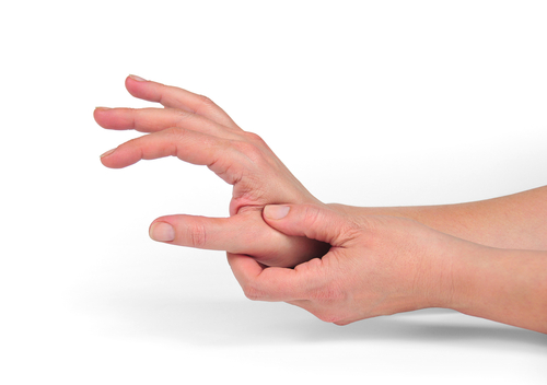
The joint capsule of the thumb mp joint is a stout structure that attaches circumferentially around the joint and seals the joint. The mp joint of the thumb is the middle joint of the thumb located between the cmc joint and the tip of the thumb.
Hand Anatomy Midwest Bone Joint Institute Elgin Illinois
The dip joint is located at the tip of the finger.

Thumb joint anatomy. The ip joint in thumb is located at the tip of the finger just before the fingernail starts. A second hole is made dorsally to volarly 1 cm distal to the base of the index metacarpal. Using wire suture or a tendon passer the slip of the apl is passed through the base of the thumb metacarpal and then volarly to dorsally in the index metacarpal.
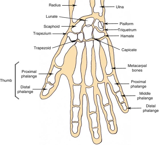
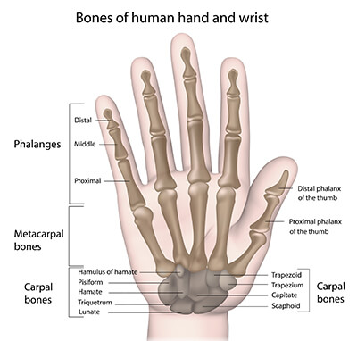
Interphalangeal joint ip the thumb digit has only two phalanges bones so it only has one joint. Distal interphalangeal joint dip. Finger movement is controlled by muscles in the forearms that pull on finger tendons.
Each of the fingers has three joints. Break down the words in the name metacarpophalangeal and you get metacarpo hand bone and phalangeal finger bone. Tendons connect muscles to bones.
The joint capsule blends with the palmar plate and the collateral ligaments. Proximal interphalangeal joint pip the joint in the middle of the finger. There are two interphalangeal joints ip joints on each finger.
Joint capsule of the thumb. The finger bones are known as phalanges singular phalanx. Fingers have a complex anatomy.
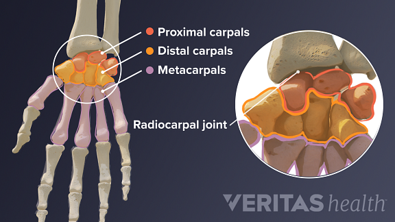
Metacarpophalangeal joint mcp the joint at the base of the finger. Distal interphalangeal joint dip the joint closest to the fingertip. Ask a doctor online now.
They are responsible for reinforcing the thumb. There are nine interphalangeal joints in each hand two on each finger and one in the thumb. The collateral ligaments are called the anterior and posterior ligaments.
The thumb interphalangeal ip joint is similar to the distal interphalangeal dip joint in the fingers. Ligaments connect finger bones and help keep them in place. The thumb has two of each.
This joint moves a lot in some people and just a little in other people. The pip joint is the joint just below the dip joint. Nerves send signals from the brain to the.
Anatomy of the fingers finger bones. The thumb joint has two collateral ligaments as well as the capsule which is lined by a synovial membrane. Proximal interphalangeal joint pip.
Nerves of the fingers. Each finger has 3 phalanges bones and 3 hinged joints. The small ringer middle and index fingers all have the same four joints.
 Motions Of The Thumb Everything You Need To Know Dr Nabil Ebraheim
Patient Education Concord Orthopaedics
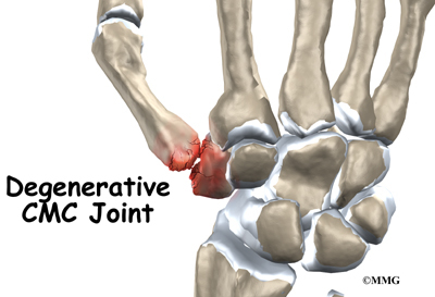
 Base Of The Thumb Arthritis 1st Cmc Arthritis Treatment
 Common Hand And Wrist Conditions Pro Sports Orthopedics
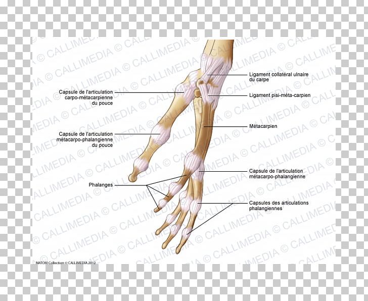 Thumb Joint Capsule Hand Human Anatomy Png Clipart Abdomen
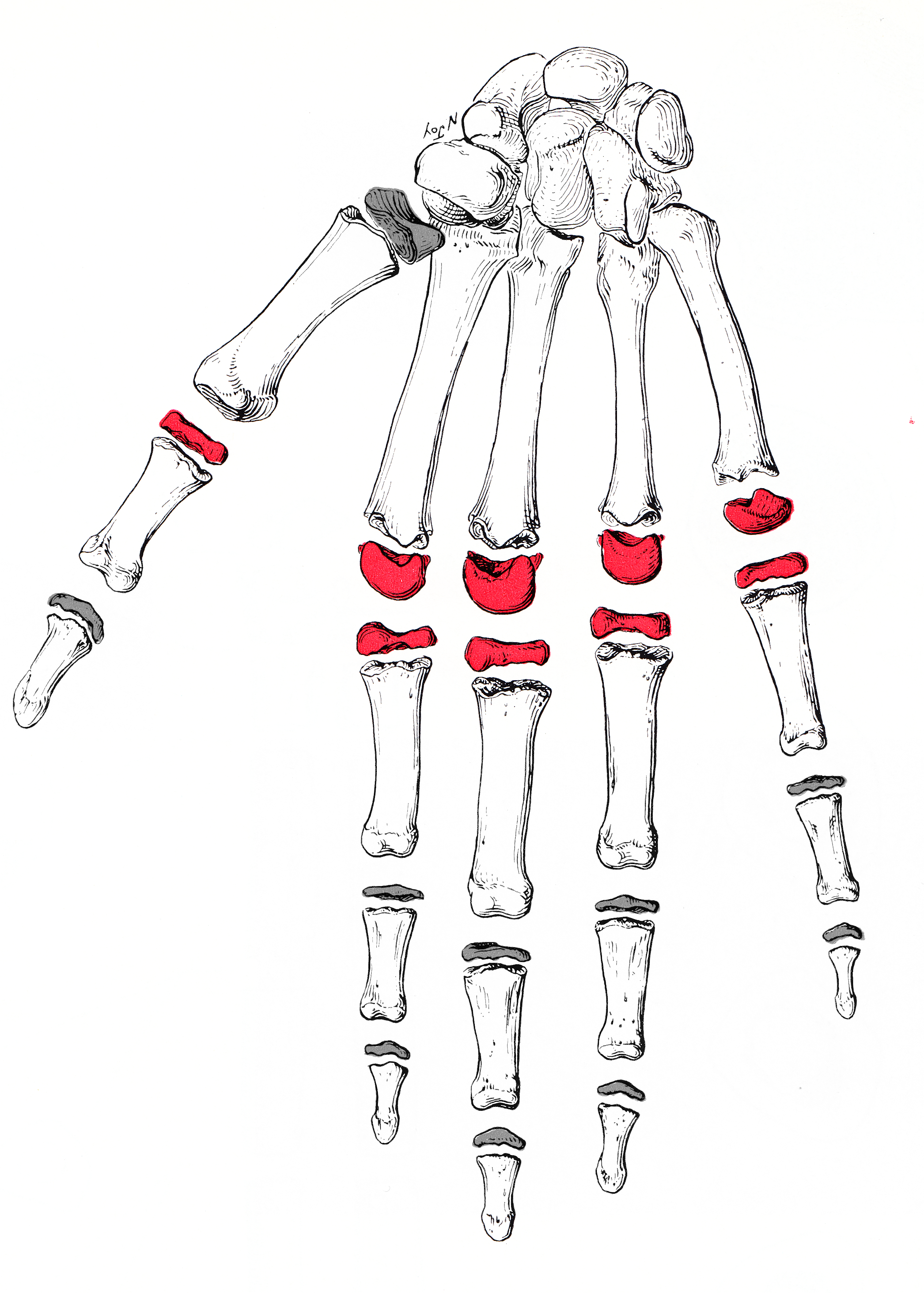 Metacarpophalangeal Joint Wikipedia
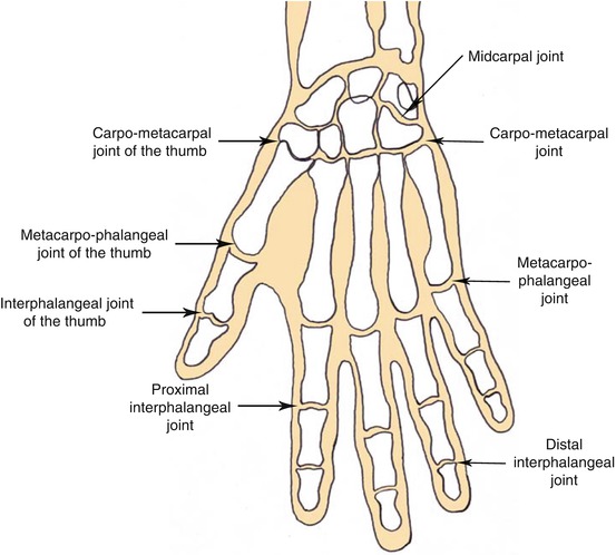 The Anatomy Of The Hand Musculoskeletal Key
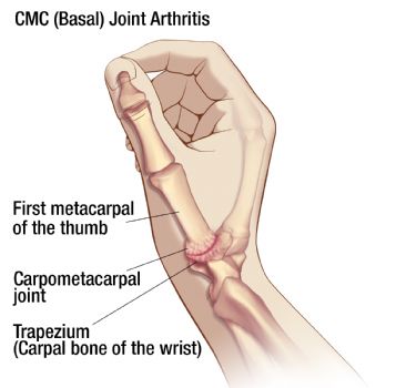
 Evaluation And Diagnosis Of Wrist Pain A Case Based
 Thumb Injuries Your Complete Guide To Diagnosing Thumb Pain
 Trigger Finger Causes Symptoms Diagnosis Treatment
 Finger Anatomy Picture Image On Medicinenet Com
Thumb Cmc Arthroplasty Thumb Joint Reconstruction
 The Hand Advanced Anatomy 2nd Ed
 Thumb Cmc Basal Joint Arthroplasty Thumb Joint
 Common Problems Of The Hand Wrist Wilmington Health
 Thumb Joint Wrist Radius Upper Limb Axillary Anatomy Png
 The Anatomy Of The Hand Musculoskeletal Key
 Cmccarethumb Brace Application Oma
 Anatomy And Biomechanics Of The Thumb Carpometacarpal Joint
 Hand Wrist Preservation Baltimore Md Towson Orthopaedics
 Interphalangeal Joint An Overview Sciencedirect Topics
 How To Treat Arthritis In The Hands Uchicago Medicine
Hand Anatomy Midwest Bone Joint Institute Elgin Illinois
Adult Thumb Metacarpal Fractures Midwest Bone And Joint


Belum ada Komentar untuk "Thumb Joint Anatomy"
Posting Komentar