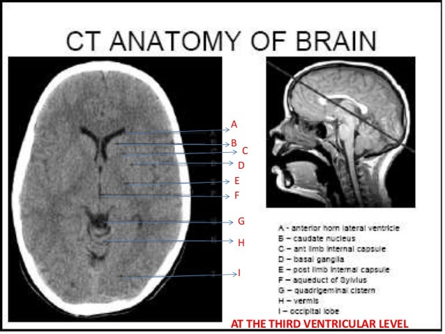
Ct Anatomy Brain
6 frontal bone 27 occipital bone 32 optic nerve 37 basilar artery 40 hemisphere of cerebellum 43 frontal sinus 45 sigmoid sinus 46 internal carotid artery 47 sphenoid bone 49 medulla oblongata 50 external auditory meatus 51 spinal central canal. Brain and face ct.
 Normal Anatomy Of The Brain On Ct And Mri With A Few Normal
Cross sectionnal anatomy of the head on a cranial ct scan.

Ct anatomy brain. Coronal brain ct. Head ct anatomy normal anatomy 1. The anterior part of the head is at the top of the image.
Neck ct cervical lymph node levels. To load the neck ct anatomy module in a new window click on its image above. Non contrast sagittal ct head.
Angiogram axial ct head. Anatomy of the head on a cranial ct scan. Interactive anatomy atlas.
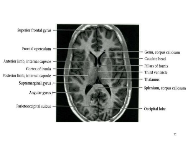
Basal forebrain on ct and mr images the basal forebrain is a rather featureless region on the ventral surface of the brain. This means that the right side of the brain is on the left side of the viewer. Non contrast coronal ct head.
Head and neck atlas. Non contrast axial ct head. Hnbs neuroanatomy modules neck ct.
Spl head and neck atlas 2012 november. This article lists a series of labeled imaging anatomy cases by system and modality. Ct images of the brain are conventionally viewed from below as if looking up into the top of the head.
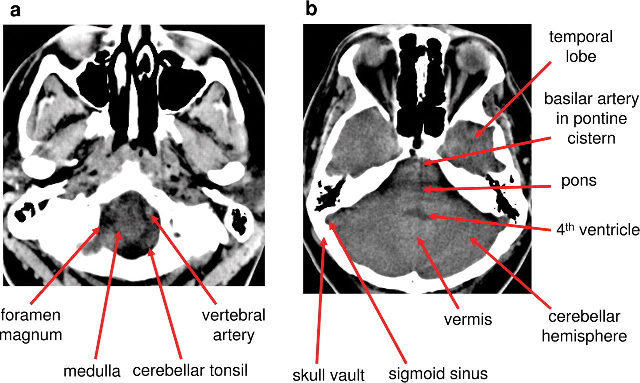
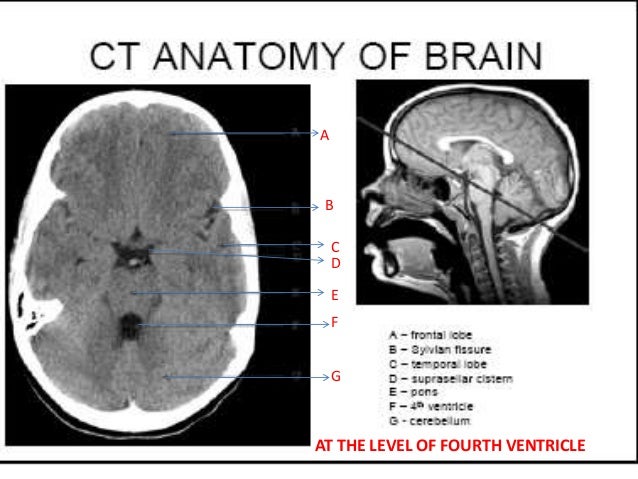
Anatomy ct axial brain form no 18. Angiogram coronal ct head. Normal ct brain of a 35 year old for reference brainstem and cerebellum without evidence of focal lesions.
Jakab m kikinis r. It contains both regions of gray and white matter in a heterogeneous fashion that are best appreciated on t1 and t2 weighted mr coronal images. Given that the file is large loading may take a few minutes.
Brain bones of cranium sinuses of the face. Thoracolumbar junction ct. Brain bones of skull paranasal sinuses.
Lateral ventricles of normal volume. Ct head neck atlas.
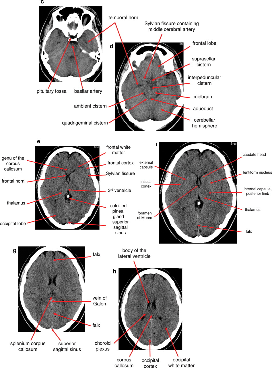
 The Radiology Assistant Brain Anatomy
 Brain Anatomy On Ct Axial Landmarks Normal Brain Ct Thi
 Normal Anatomy Of The Brain On Ct And Mri With A Few Normal
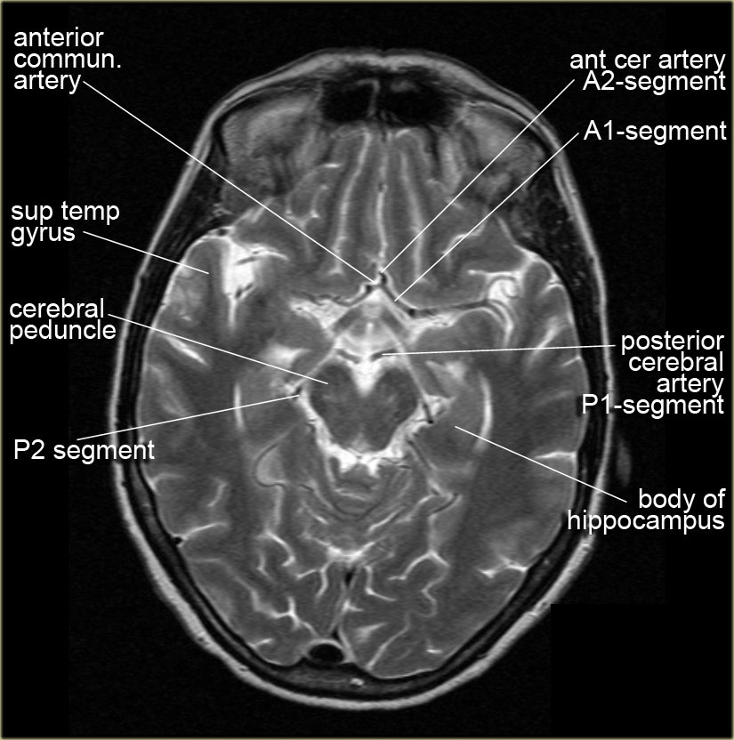 The Radiology Assistant Brain Anatomy
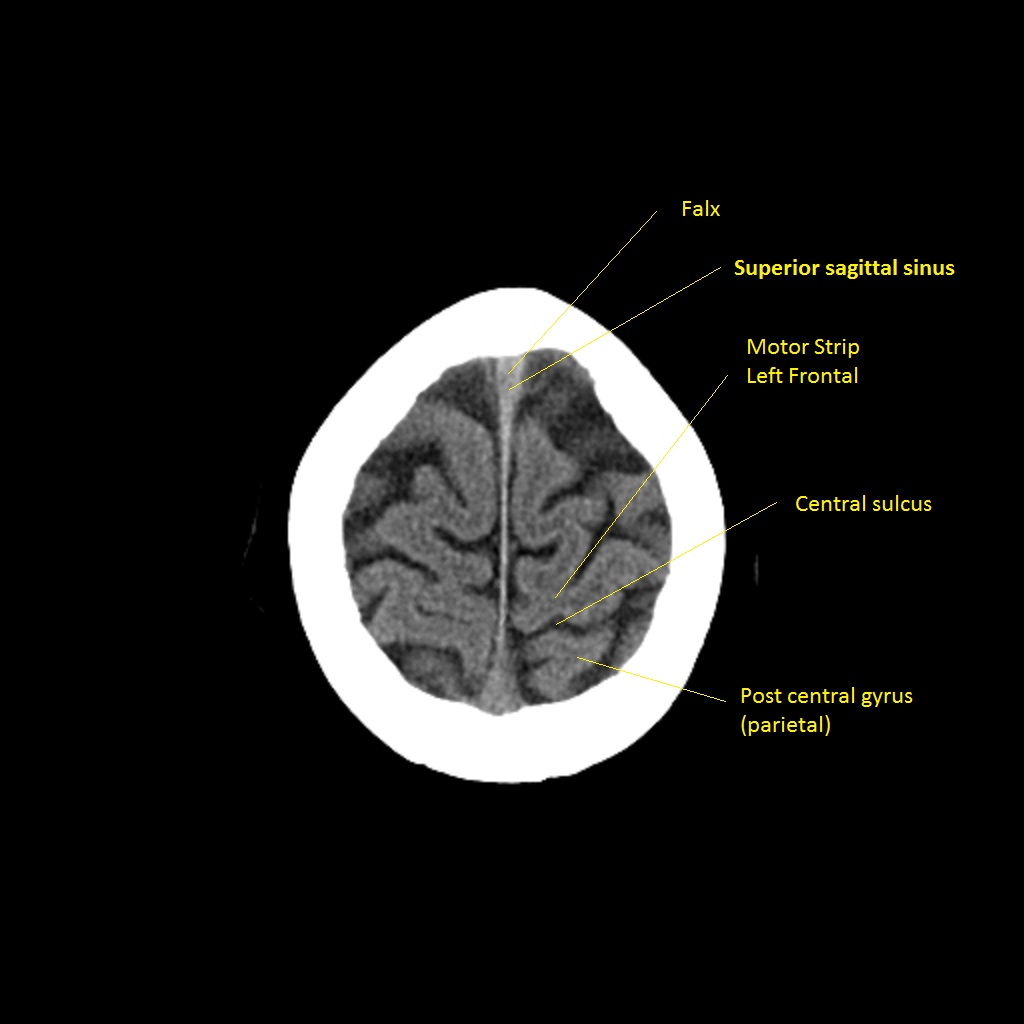
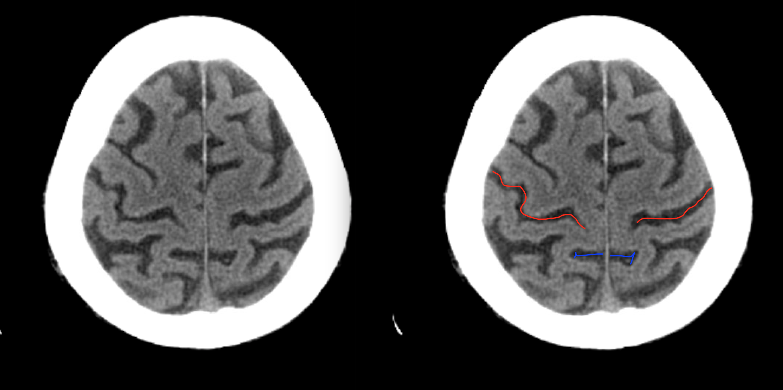 Radiological Anatomy Central Sulcus Stepwards
 Normal Ct Brain
 Ct Brain Anatomy Basal Ganglia Google Search Mri Brain
 Brain Lobes Annotated Mri Radiology Case Radiopaedia Org
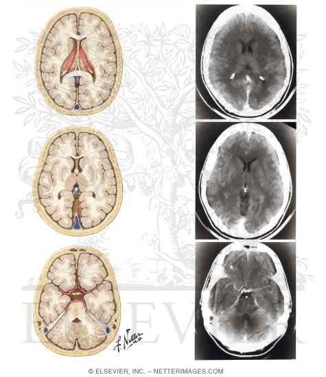 Normal Brain Anatomy As Demonstrated By Computerized
 Normal Brain Anatomy As Demonstrated By Computerized
 Stroke Medicine For Stroke Physicians And Neurologists
 Radiology Basics Head Anatomy
 Imaging Of The Central Nervous System Clinical Gate
 Brain Ct Neurologyneeds Com
 Cerebral Toxoplasmosis Infection Stock Photo Image Of
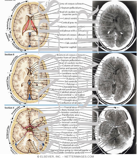
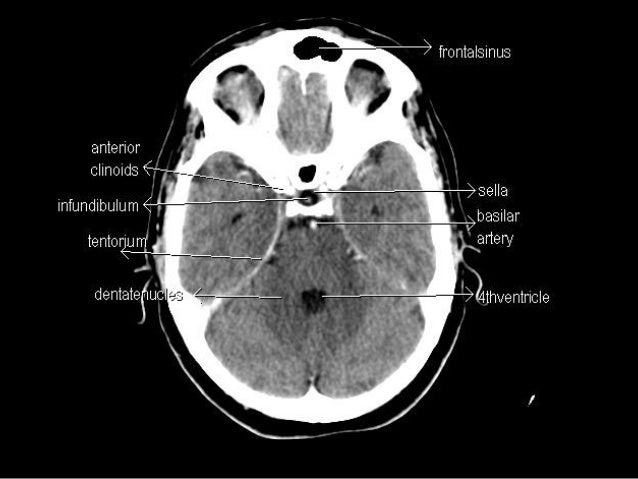 Ct Anatomy
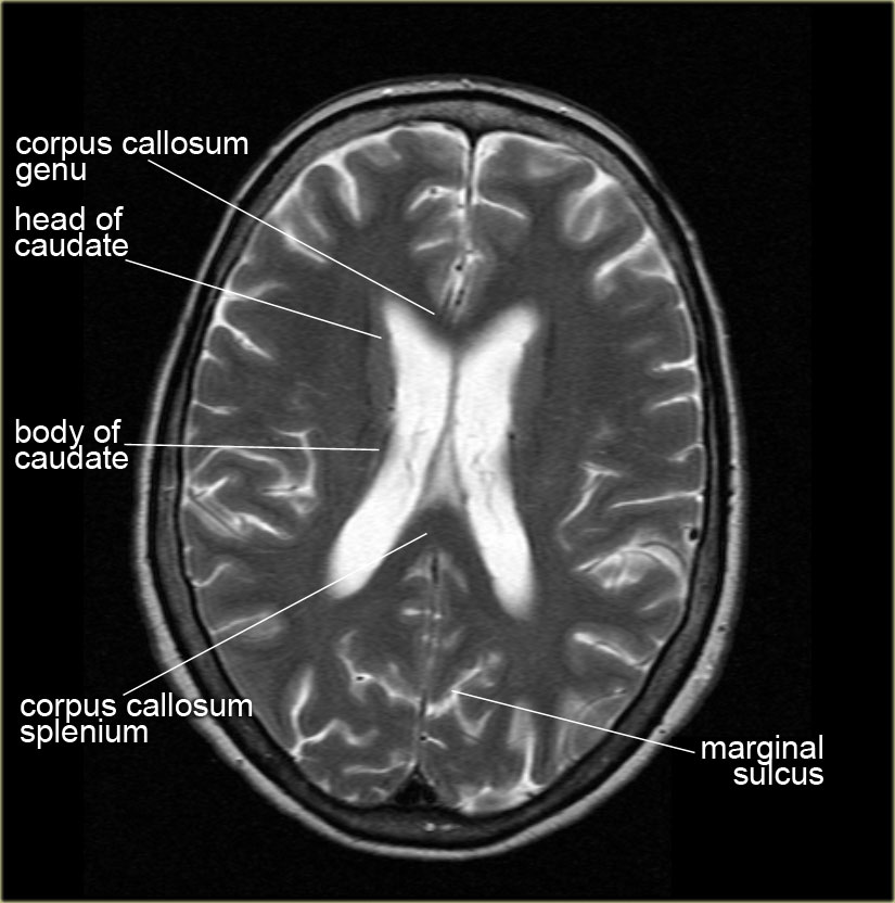
 Brain Anatomy Mri Coronal Brain Anatomy Free Mri Cross
 How To Interpret An Unenhanced Ct Brain Scan Part 1 Basic
 Headneckbrainspine
 Ct Head Interpretation Android Apps Appagg
 Paranasal Sinuses Ct Frontal Sinus Maxillary Sinus
 Ct Brain For Android Apk Download
 Ct Axial Image Of The Brain Showing The Length Of Right And
 Normal Ct Brain
 Normal Anatomy Of Brain On Ct And Mri
 Introduction To Brain Surface Anatomy
Normal Anatomy Radiology Key
 The Ct Anatomy Tutor
 The Ct Anatomy Tutor
 Figure 69 3 From How To Read A Head Ct Scan Semantic Scholar
 Ai Detects Tiny Brain Hemorrhages On Ct Scans Outperforms
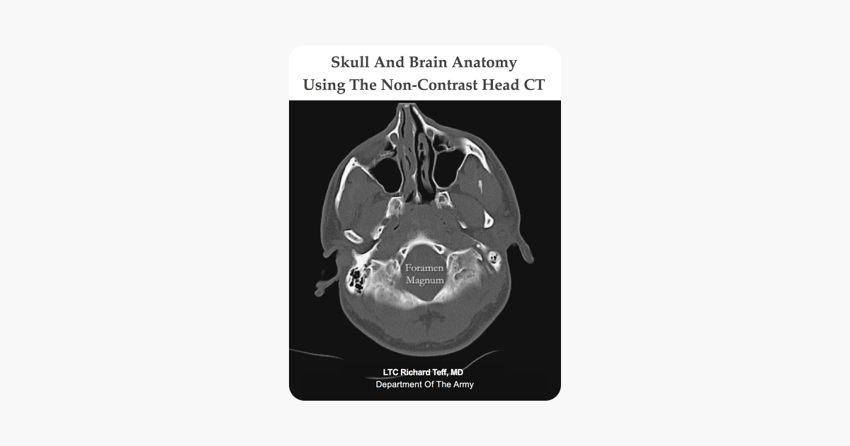 Skull And Brain Anatomy Using The Non Contrast Head Ct
 Brain Ct Neurologyneeds Com
 The Radiology Assistant Brain Anatomy


Belum ada Komentar untuk "Ct Anatomy Brain"
Posting Komentar