Peroneal Nerve Anatomy
It can cause neuropathy. The deep peroneal nerve and the superficial peroneal nerve.
Superficial Fibular Nerve Superficial Peroneal Nerve
It divides at the knee into two terminal branches.

Peroneal nerve anatomy. Schematic drawing of a sagittal a and coronal b view with corresponding axial t1 wi of a right knee. The common fibular nerve also known as the common peroneal nerve is one of two main muscular branches of the sciatic nerve. The common peroneal nerve is the smaller and terminal branch of the sciatic nerve which is composed of the posterior divisions of l4 5 s1 2.
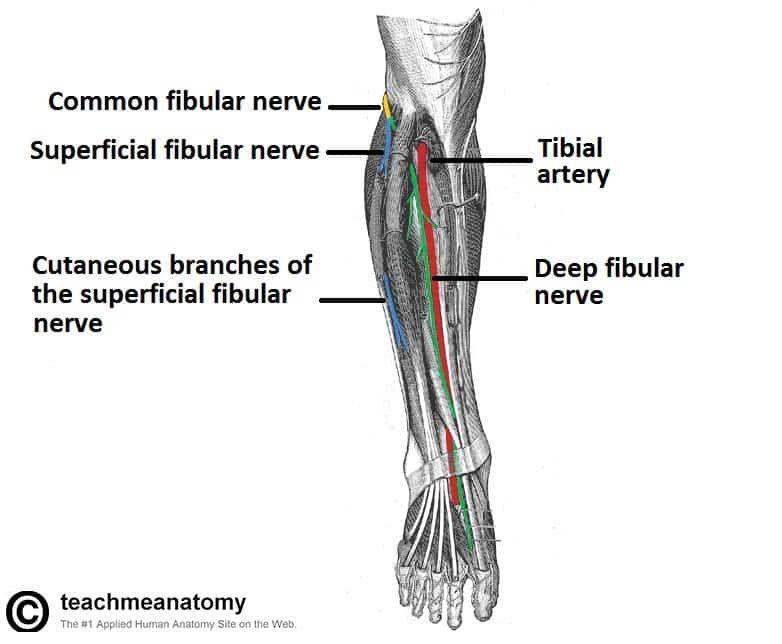
Fibularis longus fibularis brevis. Muscular branches. The superficial fibular nerve and deep fibular nerve which innervate the muscles of the lateral and anterior compartments of the leg respectively.
The common peroneal nerve is a mixed nerve it contains sensory and motor fibers. Normal anatomy of the peroneal nerve at the level of the posterolateral corner of the right knee. The levels are indicated by the transparent boxes.
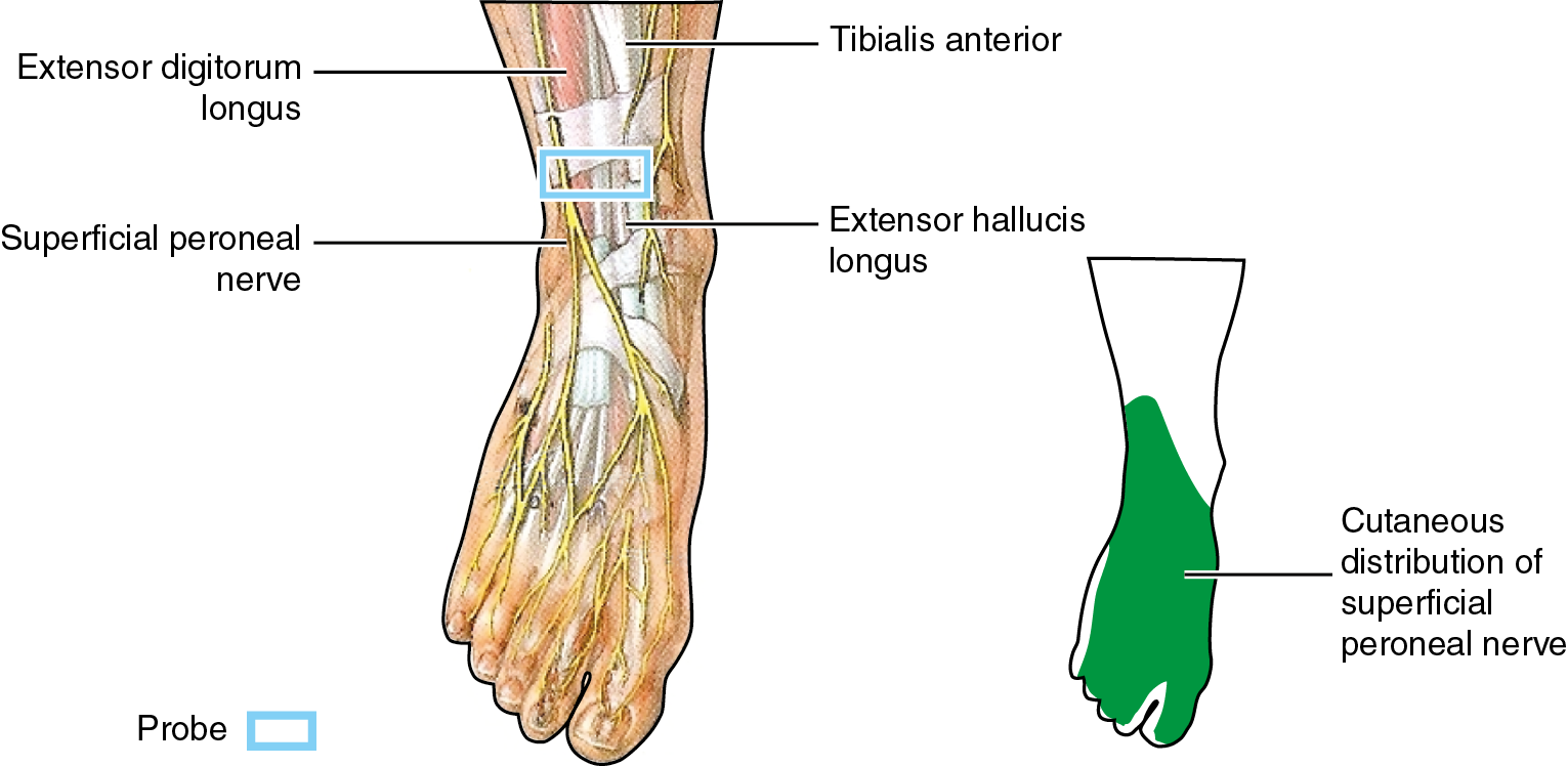
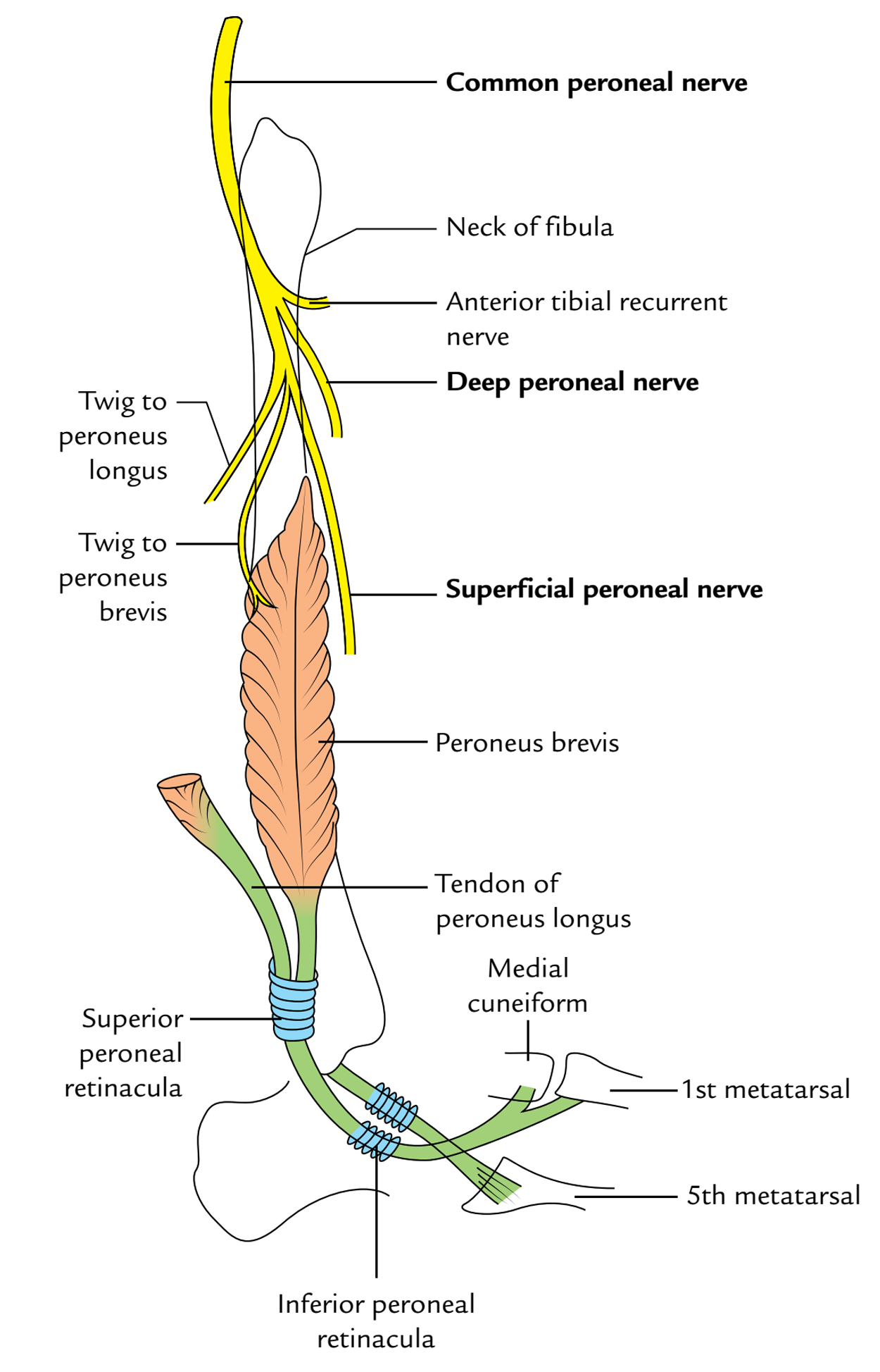
The muscles the superficial peroneal nerve innervates are. Begins at the bifurcation of the common peroneal nerve proximal neck of fibula passes between peroneal muscles and lateral side of extensor digitorum longus gives off motor branches to peroneus longus and brevis. Superior and inferior genicular nerves travel with the arteries of the same name and supply.
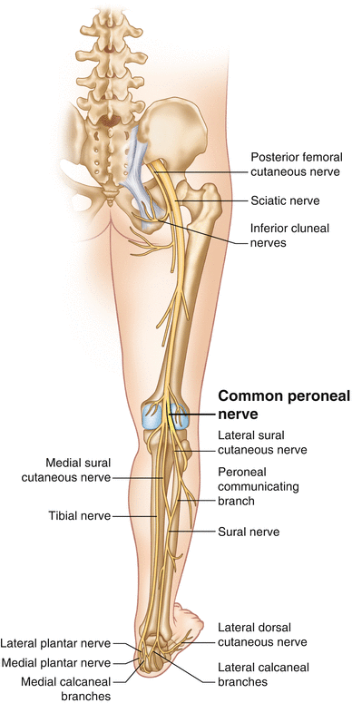
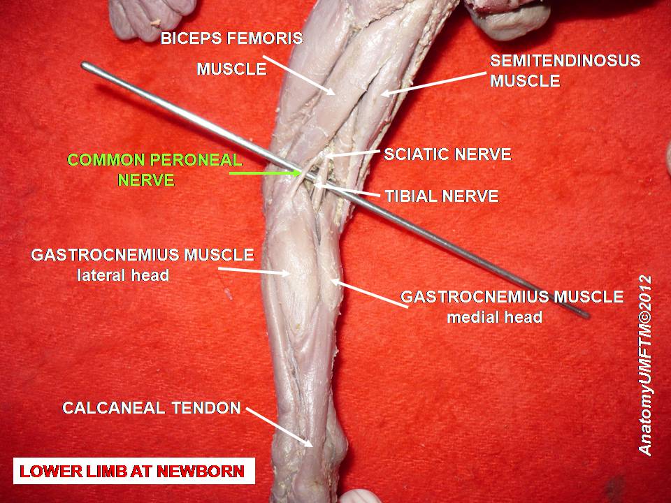
The common peroneal nerve starts just above the knee and supplies sensory and motor function to the lower leg and foot. It courses along the upper lateral side of the popliteal fossa deep to biceps femoris and its tendon until it gets to the posterior part of the head of the fibula. The common fibular nerve is a nerve in the lower leg that provides sensation over the posterolateral part of the leg and the knee joint.
The common peroneal nerve arises above the popliteal fossa runs along the medial edge of the biceps femoris to the neck of the fibula where it divides into terminal branches. This common fibular nerve then divides into the deep and superficial fibular nerves and innervates the muscles listed. Branches and supply peroneal communicating nerve.
When the common fibular nerve is damaged or compressed foot drop can be the end result. Lateral sural cutaneous nerve pierces the roof of the fossa over the lateral gastrocnemius head.

 48 Deep Peroneal Nerve Anterior Tibial Nerve
 Superficial Peroneal Nerve Sonoanatomy For Anaesthetists
 Sciatic Nerve An Overview Sciencedirect Topics
 Superficial Peroneal Nerve Anatomy Orthobullets
 Common Peroneal Nerve Seriously Sciatic
 An Uncommon Case Of Bilateral Peroneal Nerve Palsy Following
 Common Fibular Nerve An Overview Sciencedirect Topics

 Locating The Anterior Common Peroneal Nerve Neural Surface Anatomy Series Stimpod Nms460
 The Common Fibular Nerve Course Motor Sensory
 Easy Notes On Superficial Peroneal Nerve Learn In Just 3
 Common Peroneal Nerve Entrapment Springerlink
 The Common Fibular Nerve Course Motor Sensory
 Cutaneous Nerve Blocks Of The Lower Extremity Nysora
Accessphysiotherapy Lumbar And Sacral Plexus With Clinical
 Entrapment Neuropathies In The Upper And Lower Limbs
 Peroneal Nerve Paralysis Wikipedia
 Ao Surgery Reference
 Superficial Peroneal Nerve Anatomy Orthobullets
 The 4 Main Patterns Of Superficial Peroneal Nerve Anatomy
 Common Peroneal Nerve Nerve Anatomy Compartment Syndrome
 Peripheral Nerve Injury Map Axogen
![]() Common Fibular Nerve Peroneal Anatomy And Innervation
Common Fibular Nerve Peroneal Anatomy And Innervation
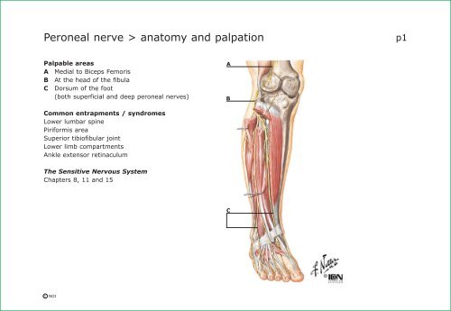 Peroneal Nerve Anatomy And Palpation

 Common Peroneal Nerve Neurologyneeds Com
 Common Peroneal Nerve Peroneus Longus Foot Superficial
Belum ada Komentar untuk "Peroneal Nerve Anatomy"
Posting Komentar