Ct Temporal Bone Anatomy
Each temporal bone is composed of five osseous parts. Recent advances in 32 64 and now 128 slice ct scanners allow the acquisition of high resolution volumetric data that allows image reconstruction in any plane.
 Ct Scan Of The Temporal Bone Overview Normal Anatomy Of
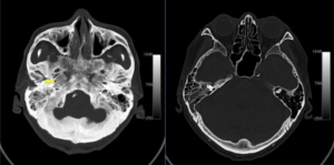
In this review we present the normal axial and coronal anatomy of the temporal bone by scrolling through the images.

Ct temporal bone anatomy. The temporal bone is situated on the sides and the base of the cranium and lateral to the temporal lobe of the cerebrum. Given that the file is large loading may take a few minutes. The temporal bones comprise the lateral skull base forming portions of the middle and posterior fossae.
Ct is the imaging modality of choice for most of the pathologic conditions of the temporal bone especially for those of the middle ear. You will find more temporal bone pathology here. The squamous mastoid petrous tympanic and styloid portions.
This atlas allows you to scroll through ct slices of the temporal bone in four different planes. Ct scan of the temporal bone. Disease processes in the pontine angle and in the internal acoustic meatus are not discussed.
Click on an image to select a plane. To load the temporal bone ct anatomy module in a new window click on its image above. Mri is more useful for diseases of the inner ear.
Ct scan of the temporal bone. The temporal bone is one of the most important calvarial and skull base bones. Computed tomography ct has revolutionized imaging of the temporal bone.
The temporal bone is very complex and consists of five parts. The module interface is meant to mimic a radiology workstation with adjacent image scrolling via arrow keys and or mouse wheel button. Temporal bone anatomy is complex and further complicated by the small size and three dimensional orientation of associated structures.
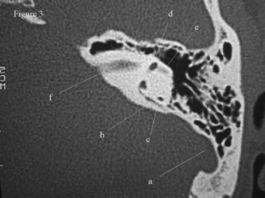
Anatomy of the petrous bone ct atlas of human anatomy using cross sectional imaging we have created an atlas of the temporal bone which is an educational tool for studying the normal anatomy of the petrous bone based on an mdct exam of the axial and coronal of the ear and petrous bone. Some structures are discussed in more detail with emphasis on related pathology. This gallery of images presents the anatomy of the temporal bone by means of ct scan reconstructions.
 Comparison Of 45 Oblique Reformats With Axial Reformats In
 The Radiology Assistant Temporal Bone Anatomy
 Temporal Bone Radiology
 Radiology Walkthroughs Ct Temporal Bone Anatomy
 Figure 2 From Imaging Of The Temporal Bone Semantic Scholar
 Normal Temporal Bone Ct Springerlink
 Inflammatory Ear Conditions
 Temporal Bone Radiology Reference Article Radiopaedia Org
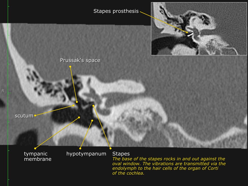 The Radiology Assistant Temporal Bone Anatomy 2 0
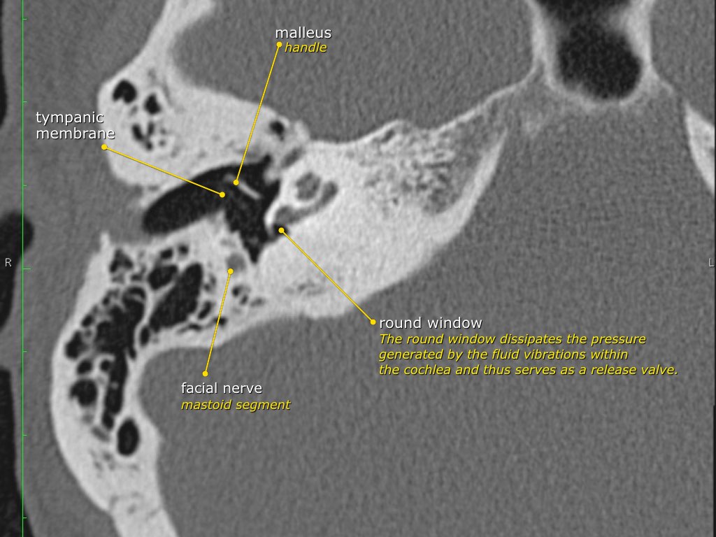 The Radiology Assistant Temporal Bone Anatomy 2 0
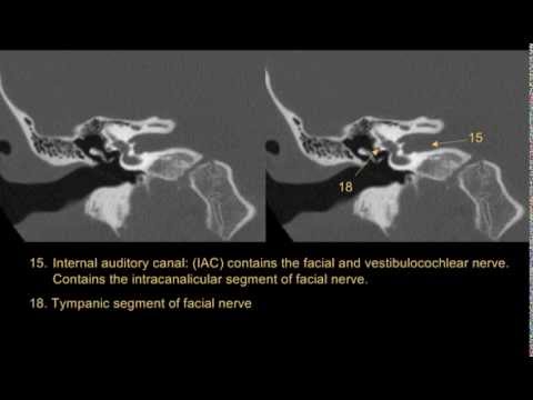 Temporal Bone Anatomy Youtube
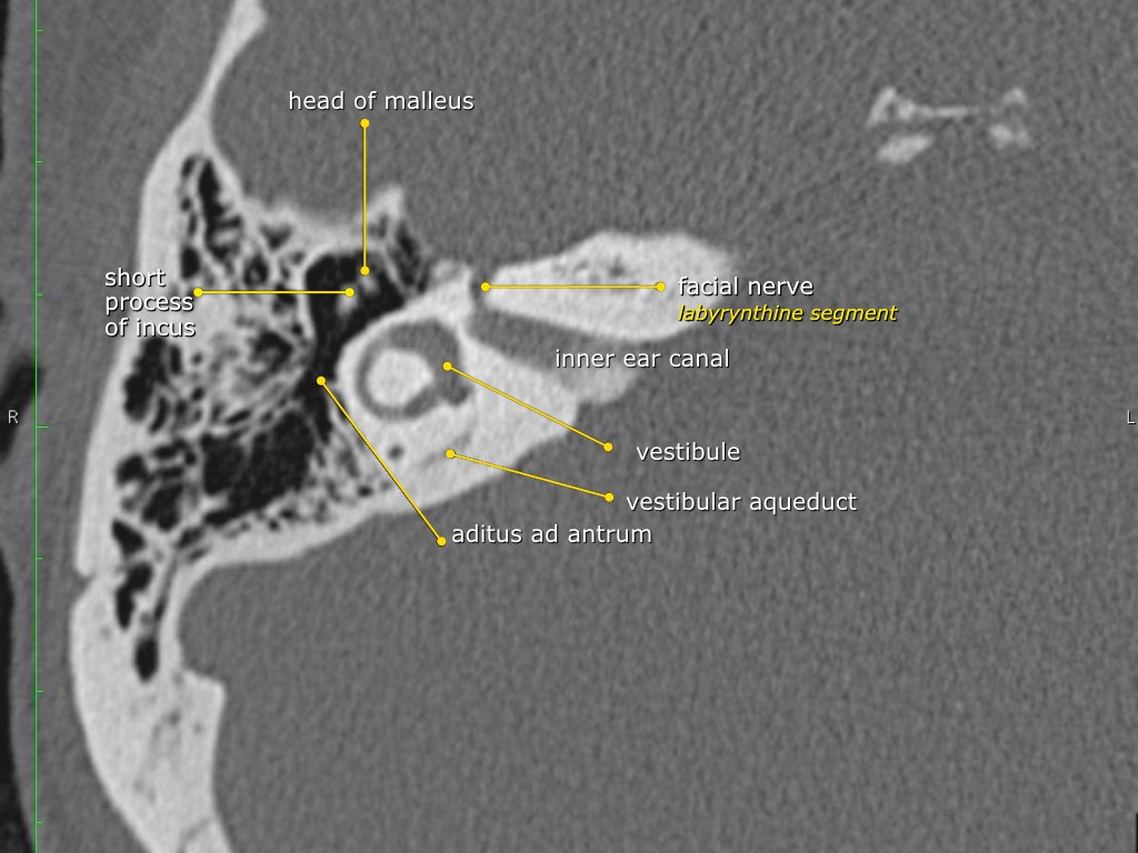
 The Radiology Assistant Temporal Bone Anatomy 2 0
 The Radiology Assistant Temporal Bone Anatomy 2 0
 Ct Scan Of The Temporal Bone
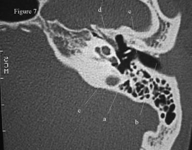
 Anatomy Of The Petrous Bone Ct
 Temporal Bone An Overview Sciencedirect Topics
 Coronal Ct Images Show The Normal Anatomy Of The Temporal
![]() Medical Imaging And Radiological Anatomy X Ray Ct Mri
Medical Imaging And Radiological Anatomy X Ray Ct Mri
 Emdocs Net Emergency Medicine Educationbasilar Skull
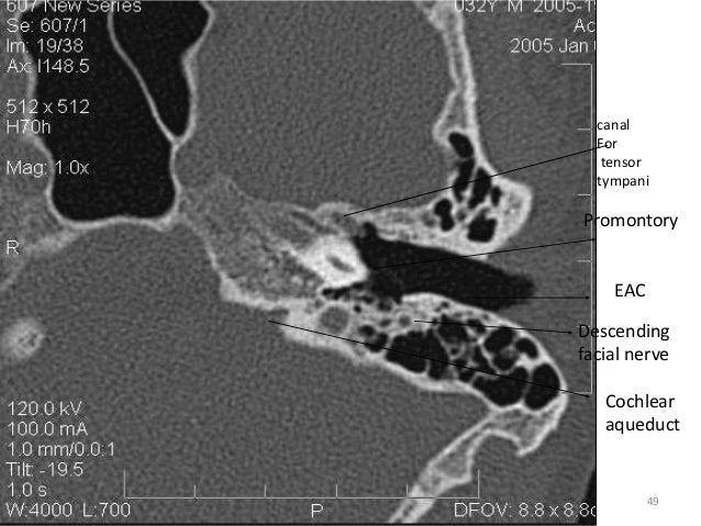
 Ppt Ct Temporal Bone Powerpoint Presentation Free
 Radiology Anatomy Images Ct Temporal Bone Anatomy
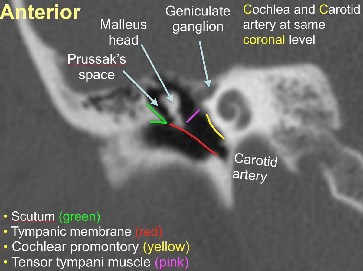 Duke Radiology On Twitter Primer On Ct Temporal Bone
 Headneckbrainspine
 Temporal Bone Radiology
 Ct Scan Of The Temporal Bone Overview Normal Anatomy Of


Belum ada Komentar untuk "Ct Temporal Bone Anatomy"
Posting Komentar