Temporal Lobe Anatomy
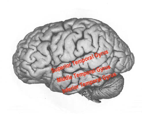
The outer surface of the temporal lobe is an association area made up of the superior middle and inferior temporal gyri. The dominant temporal lobe which is the left side in most people is involved in understanding language and learning and remembering verbal information.
 Approach To Temporal Lobe
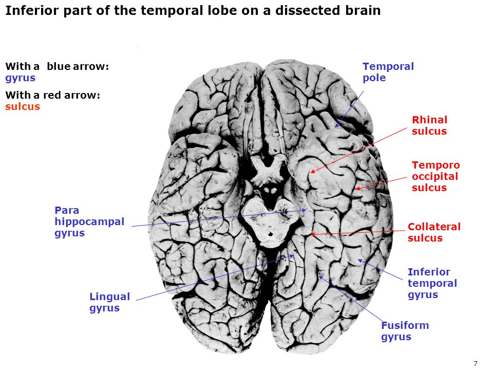
Anatomy of the temporal lobe.

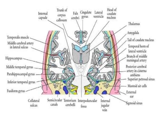
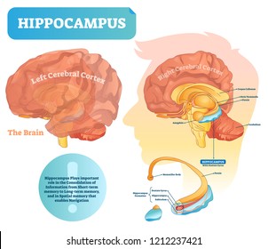
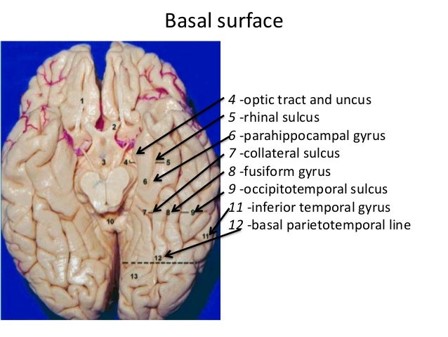
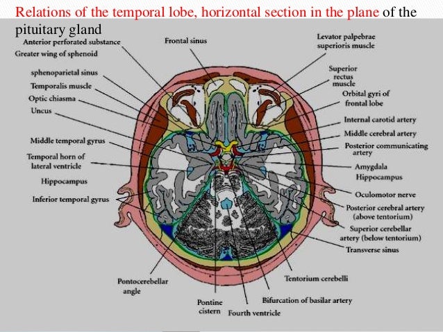
Temporal lobe anatomy. The hippocampal formation on the medial side of the lobe includes the parahippocampal gyrus subiculum hippocampus dentate gyrus and associated white matter notably the fimbria whose fibres continue into the fornix. The temporal lobe occupies the middle cranial fossa which is bounded anteriorly by the greater wing of the sphenoid bone inferiorly by the superior surface of the petrous part of the temporal bone and laterally by the squamous part of the temporal bone and the adjoining parietal bone. Structures of the limbic system including the olfactory cortex amygdala and the hippocampus are located within the temporal lobes.
The temporal lobe occupies the middle cranial fossa which is bounded anteriorly by the greater wing of the sphenoid bone inferiorly by the superior surface of the petrous part of the temporal bone and laterally by the squamous part of the temporal bone and the adjoining parietal bone. The hippocampus is an inrolled gyrus that bulges into the temporal horn of the lateral ventricle. Gross anatomy the temporal lobe is the second largest lobe after the large.
Structures of the cerebral cortex. The temporal lobe is crucial in many essential activities such as processing of memory language and emotion. The temporal lobe located just beneath the lateral fissure and crisscrossing both fissures of the brain.
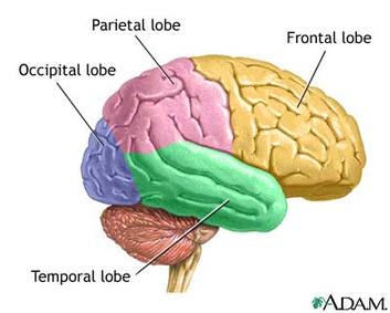
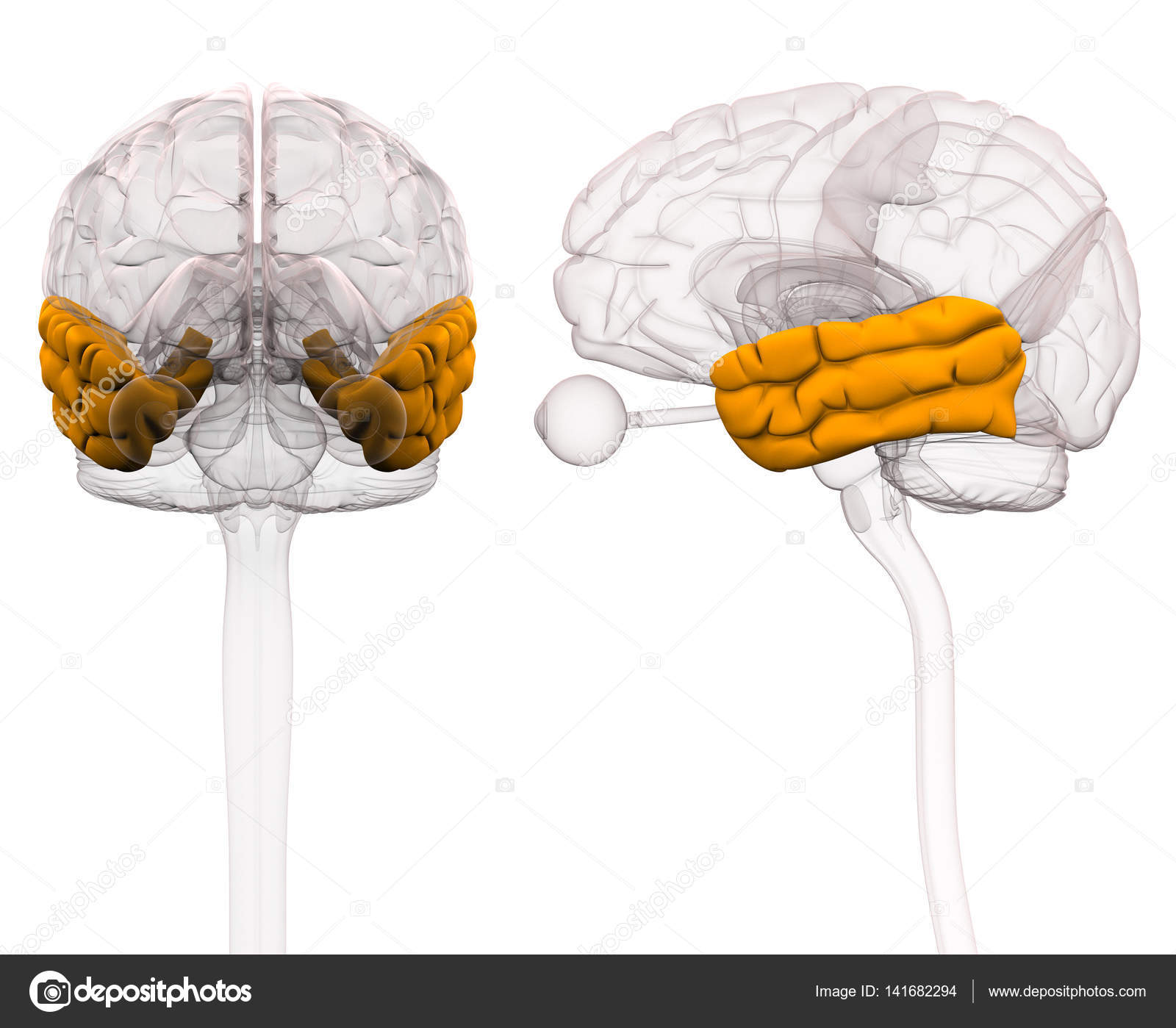
The temporal lobe is one of the four lobes of the brain along with the frontal lobe parietal lobe and occipital lobe and largely occupies the middle cranial fossa. The temporal lobe is involved in processing sensory input into derived meanings for the appropriate retention of visual memory language comprehension and emotion association. Gross anatomy the temporal lobe is the second largest lobe after the large.
This vital structure of the temporal lobe supports process the sensory input including pain and the auditory stimuli. The temporal lobe is located beneath the lateral fissure on both cerebral hemispheres of the mammalian brain. In human nervous system.
Damage to this area of the brain can result in problems with memory understanding language and maintaining emotional control. The temporal lobes play an important role in organizing sensory input auditory perception language and speech production as well as memory association and formation. Lobes of the cerebral cortex the temporal lobe inferior to the lateral sulcus fills the middle fossa or hollow area of the skull.
The brains contain four lobes in the cortex including the occipital parietal temporal and frontal lobes. The temporal lobe is one of the four lobes of the brain along with the frontal lobe parietal lobe and occipital lobe and largely occupies the middle cranial fossa.
 How To Delineate The Different Parts Of The Temporal Lobe
 Temporal Lobe Functions And Syndromes Psy
 Memory Part 2 The Role Of The Medial Temporal Lobe
 Occipital Lobe Radiology Reference Article Radiopaedia Org
 The Lobes Human Brain
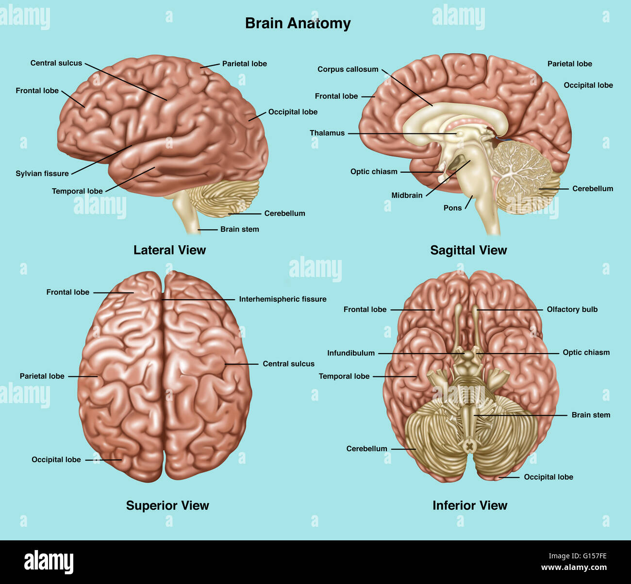 Illustration Showing Anatomy Of A Normal Brain In Lateral
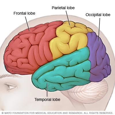 Brain Lobes Mayo Clinic
 Cognitive Skills Of The Brain Brain Injury Alliance Of Utah
 Temporal Lobe Human Brain Series Part 8
![]() Lobes Of The Brain Anatomy Functions And Clinical Facts
Lobes Of The Brain Anatomy Functions And Clinical Facts
Anatomical Relations Of The Temporal Lobe As Seen In A
 Inferior Temporal Gyrus Wikipedia
 Temporal Lobe Images Stock Photos Vectors Shutterstock
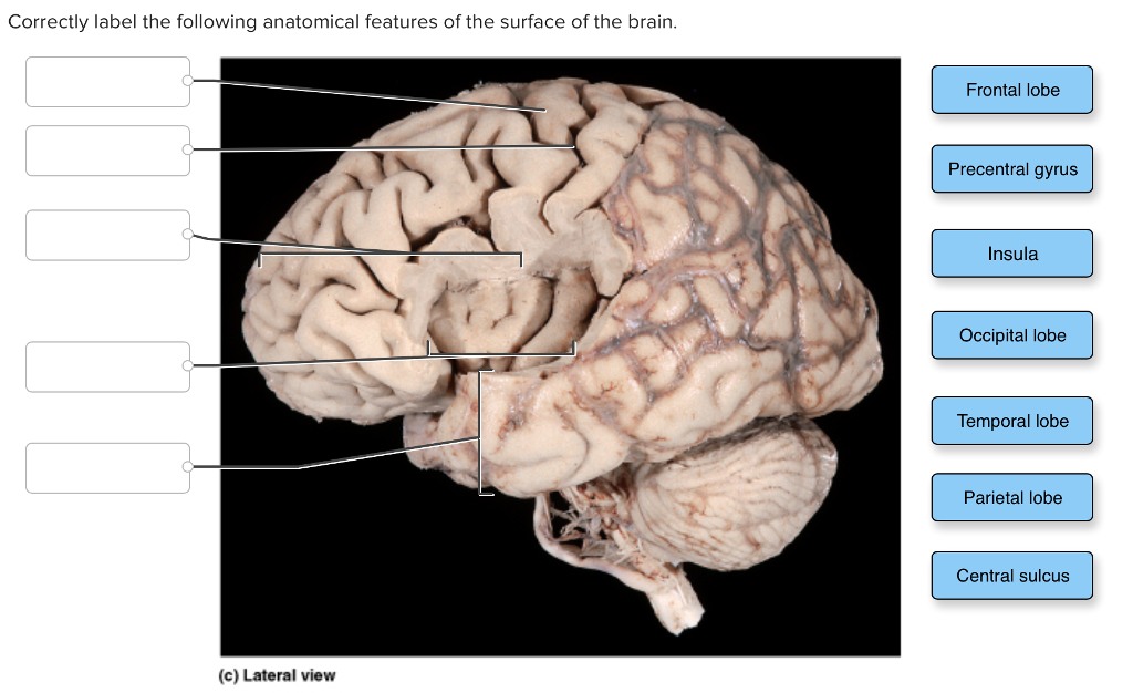 Solved Correctly Label The Following Anatomical Features
 Brain Anatomy Temporal Lobe
 Temporal Brain Lobe Position Structure Function Role
 Brain Anatomy Temporal Lobe Art Print Poster
 The Brain
 Brain Structure And Function
 Location Of The Temporal Lobes It Is On Both Sides Behind
 Medial Temporal Lobe An Overview Sciencedirect Topics
 Final Microsurgical Anatomy Of Medial Temporal Lobe
 Approach To Temporal Lobe Anatomy Function Epilepsy Mri Finding
 Temporal Lobe
 Brain Part C Frontal Lobe Temporal Lobe Parietal Lobe
 4 A Illustration Showing The Location Of Medial Temporal
 Boundaries Of The Temporal Lobe And Positions Of Major Sulci
 Temporal Lobe Brain Anatomy 3d Illustration Stock Photo

Belum ada Komentar untuk "Temporal Lobe Anatomy"
Posting Komentar