Lateral Ventricle Anatomy
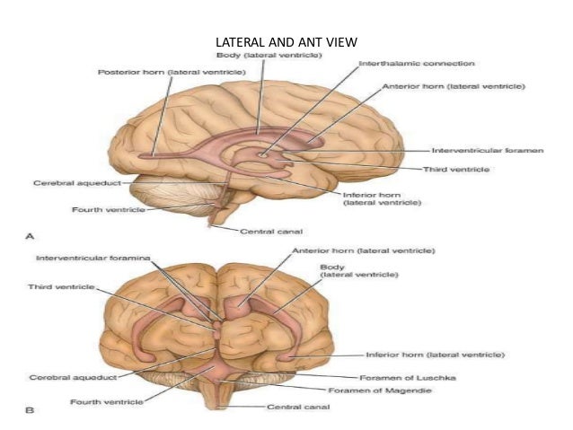
The lateral ventricles are c shaped. Each is divided into a central part composed of the body and trine or atrium and three lateral extensions orhorns.
 View Large Accessneurology Mcgraw Hill Medical
The lateral ventricles are paired structures and part of the ventricular system in the brain.
Lateral ventricle anatomy. These five divisions are the anterior frontal horn body. The volume of the lateral ventricles increases with age. From the central part three extensions are given which are as follows.
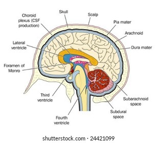
It is the classic location for intraventricular meningioma. The trigone of the lateral ventricle also known as the atrium is a triangular area at the floor of the lateral ventricle that forms the transition point between the occipital and temporal horns. The lateral ventricles are connected to the third ventricle by the foramen of monro.
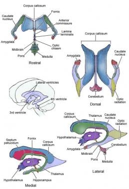
Each cerebral hemisphere contains a lateral ventricle known as the left or right ventricle respectively. The right and left lateral ventricles are structures within the brain that contain cerebrospinal fluid a clear watery fluid that provides cushioning for the brain while also helping to circulate nutrients and remove waste. It is located in the parietal lobe of cerebrum.
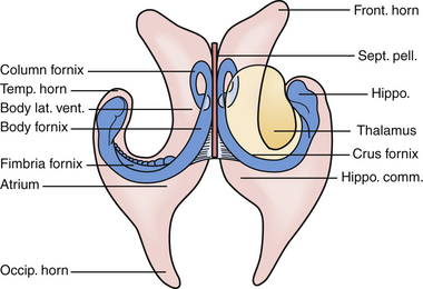
Anatomically the lateral ventricle can be thought of as a c shaped capsule encompassing the thalamus and diencephalon. The anterior horn of the lateral ventricle lies anterior to the central part. The lateral ventricles are the two largest cavities of the ventricular system of the human brain and contain cerebrospinal fluid csf.
The third ventricle is situated in between the right and the left thalamus. The central part of the lateral ventricle is elongated anteroposteriorly. The central part of the lateral ventricle is called the cella media.
Each lateral ventricle is c shaped cavity divided into 4 parts. Name the parts of lateral ventricles. It is partitioned into five segmental divisions which hold important distinctions during consideration of surgical approaches.
Csf is produced in the choroid plexus situated within the ventricles and exits along the interventricular foramen of monro to the third ventricle. Anterior horn in the frontal lobe. Along with the structures known as the third ventricle and the fourth ventricle.
They are the largest cavities of the ventricular system and there is one inside each hemisphere dividing into right ventricle and left ventricle. Posterior horn in the occipital lobe inferior horn in the temporal horn. The left and right lateral ventricles are located within their respective hemispheres of the cerebrum.
They have horns which project into the frontal occipital and temporal lobes. The inferior horn is the largest component of the lateral ventricle.
 Microsurgical Anatomy Of Lateral Ventricles
 Ventricles Of The Brain Locate The Following 1 2 X
 Microsurgical Approaches To The Ventricular System
 Ventricles Of The Brain Overview Gross Anatomy

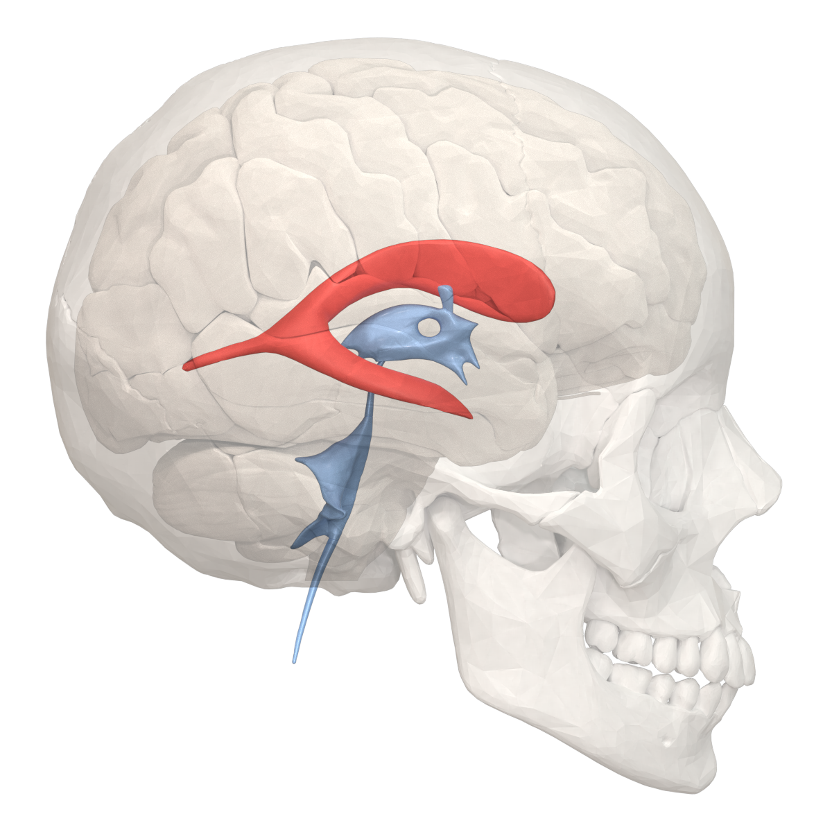 File Lateral Ventricle 02 Png Wikimedia Commons
 Lateral Ventricles An Overview Sciencedirect Topics
 Trigone Of The Lateral Ventricle Radiology Reference
 Image Result For Caudate Nucleus And Frontal Horn Of Lateral
![]() Lateral Ventricles Anatomy And Function Kenhub
Lateral Ventricles Anatomy And Function Kenhub
Occipital Horn Of Lateral Ventricle Posterior Horn Of

 Lateral Ventricle Radiology Reference Article
 M202 T1 L11 The Ventricular System Flashcards Quizlet
 Ventricular System Wikipedia
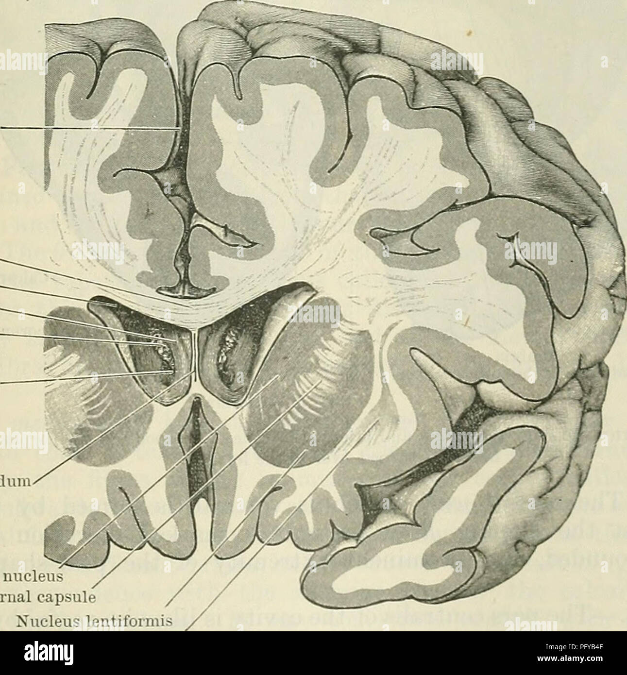 Cunningham S Text Book Of Anatomy Anatomy Longitudinal
 Lateral Ventricle Anatomy Diagram Quizlet
 Brain Ventricles Images Stock Photos Vectors Shutterstock
Lateral Ventricle Anatomy
 Ventricular System Basicmedical Key
![]() Lateral Ventricles Anatomy And Function Kenhub
Lateral Ventricles Anatomy And Function Kenhub
![]() Lateral Ventricles Anatomy And Function Kenhub
Lateral Ventricles Anatomy And Function Kenhub
 Temporal Horn Of The Lateral Ventricle Anatomical Terms Pronunciation By Kenhub
Brain Medial Child Anatomy Hp Image Details Nci
Belum ada Komentar untuk "Lateral Ventricle Anatomy"
Posting Komentar