Transitional Anatomy
Focusing on translational research the journal aims to disseminate the knowledge that is gained in the basic science of anatomy and to apply it to the diagnosis and treatment of human pathology in order to improve individual patient well being. Functions in sensing consists of the brain and spinal cord extends from the spinal cord out to the entire body.
 A Review Of Symptomatic Lumbosacral Transitional Vertebrae
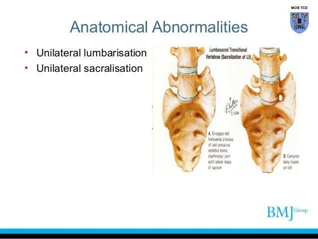
The anomalous segments are frequently referred to as transitional vertebrae lumbarization occurs due to non fusion of the first two sacral segments allowing the lumbar spine too have what appear to be six segments the sacrum appears to have only 4 segments rather than its normal 5.
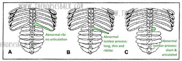
Transitional anatomy. Transitional vertebrae are abnormally formed vertebral bones that display the characteristics of two different types of vertebrae. Lumbosacral transitional vertebrae lstv are increasingly recognized as a common anatomical variant associated with altered patterns of degenerative spine changes. This review will focus on the clinical significance of lstv disruptions in normal spine biomechanics imaging techniques diagnosis and treatment.
Atlanto occipital junction atlanto occipital assimilation. There is a fairly common spinal deformity a transitional vertebra in which the lowest vertebrae of the spine is partially merged with the sacrum. They occur at the junction between spinal morphological segments.
A transitional vertebra is a somewhat common lumbosacral spinal abnormality in which one spinal bone does not form correctly and definitively as a lumbar segment or a sacral segment. Nervous covers body surfaces and lines hollow organs body cavities a protects and supports the body and its organs. Transitional vertebra is one that has indeterminate characteristic and features of vertebrae from adjacent vertebral segments.
But does not truly represent either variety in totality. Translational research in anatomy is an international peer reviewed and open access journal that publishes high quality original papers. Major communication system in the body.
It is not quite a vertebra and not quite sacrum thus transitional this study looked at how common transitional vertebrae are and if they correlate with low back pain. Complete or partial fusion of c1 and the occiput. The human spine is made up of 33 bones called vertebrae with the spinal cord running through the middle.
Instead the vertebral body takes on characteristics of both typical lumbar and sacral spinal bones. There can be a varying degree of transition from partial to complete fusion. Depending on the number of thoracic vertebrae lumbar vertebrae and sacral segments they can be thought of as a lumbarised s1 segment or sacralised l5 segment.
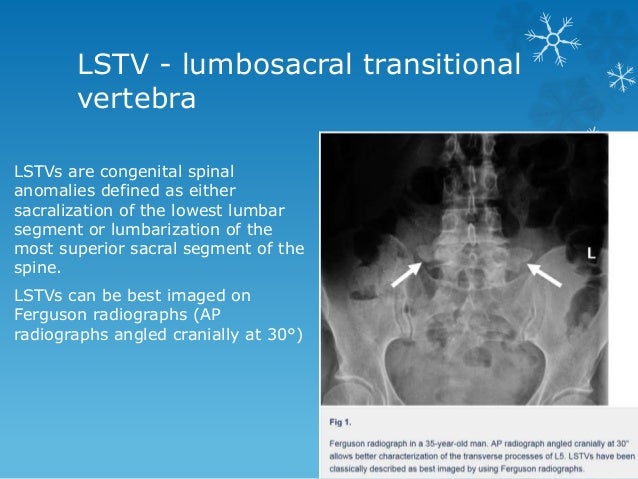 Transitional Vertebrae Radiology
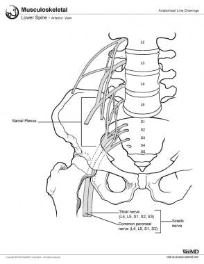 Lumbar Spine Anatomy Overview Gross Anatomy Natural Variants
 Photograph Of A Simple Sacral Dimple In A 1 Month Old Boy
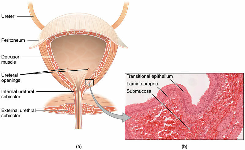 Transitional Epithelium Definition And Function Biology
Lumbosacral Transitional Vertebrae Classification Imaging
 Anatomical Differences In Patients With Lumbosacral
 An Unwelcome Side Effect Transitional Vertebrae In Horses
 Anatomical Illustration Bones Bones Brittle Little Bones
 Prediction Of Transitional Lumbosacral Anatomy On Magnetic
 Anal Canal Sphincters Introduction Anatomy
 Ureter Peritoneum Detrusor Muscle Ureteral Transitional
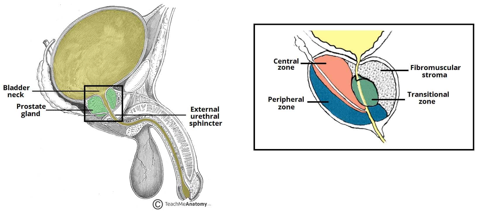 The Prostate Gland Structure Vasculature Lymph
 Anatomy Of The Spine Redlands Loma Linda Highland Bones
 It Is Composed Of Transitional Epithelium The Urinary
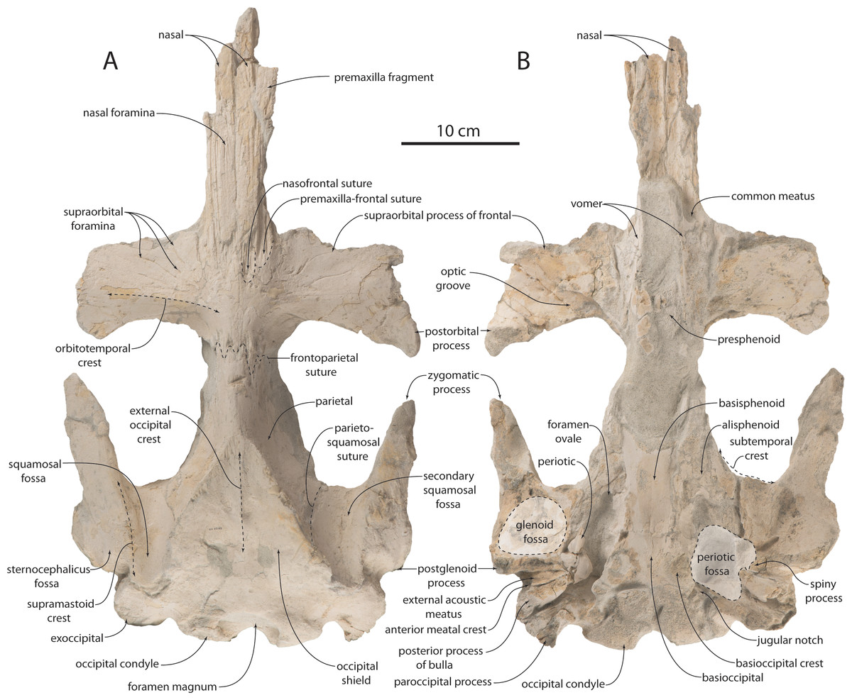 Anatomy Feeding Ecology And Ontogeny Of A Transitional
 Transitional Area Of Lower Limbs Medical Anatomy And
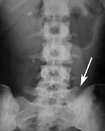 Congenital Vertebral Anomaly Wikipedia
Lumbosacral Transitional Vertebrae Classification Imaging
Lumbosacral Transitional Vertebrae Classification Imaging
 Figure 1 From Asymmetrical Lumbosacral Transitional
.png) C7 7th Cervical Vertebra Anatomy Pictures And Information
C7 7th Cervical Vertebra Anatomy Pictures And Information
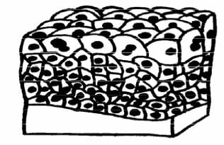 Transitional Epithelium Definition And Function Biology
Spineline November December 2017 A Case Study Of


.png)
Belum ada Komentar untuk "Transitional Anatomy"
Posting Komentar