Anatomy Of Knee Joint
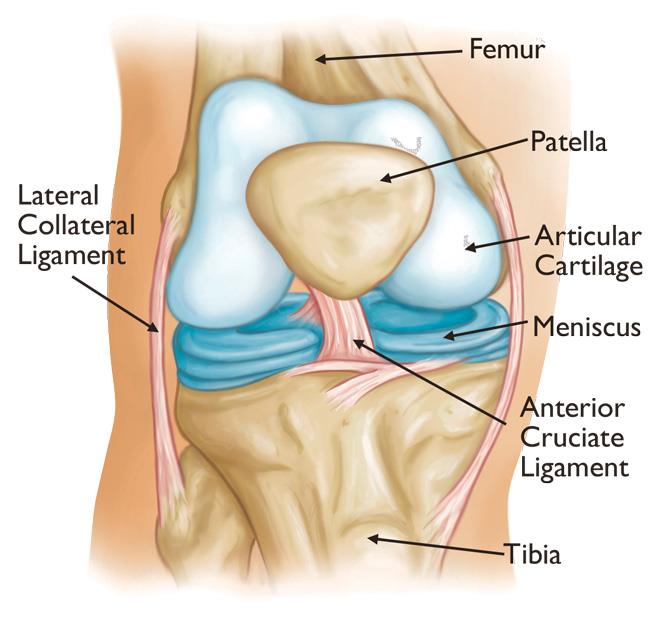
Knee joint anatomy involves looking at each of the different structures in and around the knee. Tibia the bone at the front of the lower leg or shin bone.
 Anatomy Of The Knee Baxter Regional Medical Center
The knee joins the thigh bone femur to the shin bone tibia.

Anatomy of knee joint. The knee consists of three bones. The knee is one of the largest and most complex joints in the body. Movements at the knee joint are essential to many everyday activities including walking running sitting and standing.
A hinge joint bends back and forth in one plane unlike the ball and socket joint of the hip. It is the junction of the thigh and the leg and is a hinge joint. The knee is the meeting point of the femur thigh bone in the upper leg and the tibia shinbone in the.
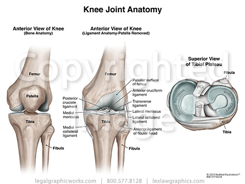
Femur the upper leg bone or thigh bone. The smaller bone that runs alongside the tibia fibula and the kneecap patella are the other bones that make the knee joint. The knee joint is a synovial joint this means it contains a fluid that lubricates it.
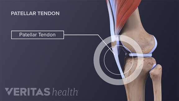
The knee joint is commonly injured. Tendons connect the knee bones to the leg muscles that move the knee joint. It allows the lower leg to move relative to the thigh while supporting the bodys weight.
This fluid is known as the synovial fluid. The knee joint is one of the strongest and most important joints in the human body. The knee joint is the largest and one of the most complex joints in the human body.
The knee joint is part of the lower extremity. The knee joint is a synovial joint which connects the femur our thigh bone and longest bone in the body to the tibia our shinbone and second longest bone. The knee is the joint where the bones of the lower and upper legs meet.
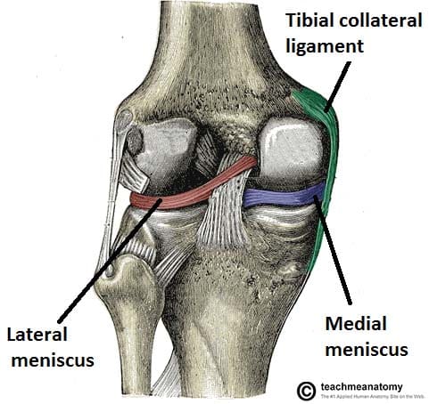
The main features of the knee anatomy include bones cartilages ligaments tendons and muscles. The knee is a complex joint that flexes extends and twists slightly from side to side. There are various muscles that control movement ligaments that give stability special cartilage to absorb pressure and various other structures to ensure smooth pain free movement.
In the knee joint the femur articulates with the tibia and the patella. The largest joint in the body the knee moves like a hinge allowing you to sit squat walk or jump. There are two joints in the kneethe tibiofemoral joint which joins the tibia to the femur and the patellofemoral joint which joins the kneecap to the femur.
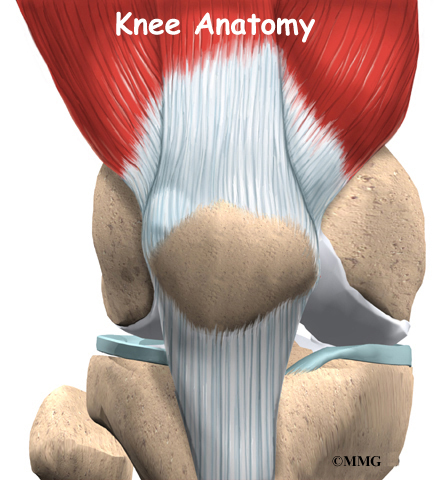
 Knee Anatomy
 Knee Joint Labelled Illustration Stock Image C043
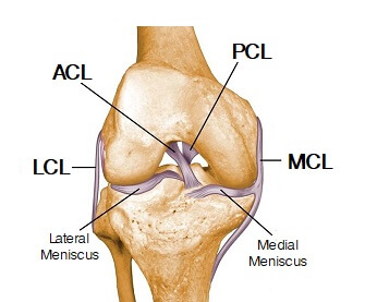 Knee Joint Anatomy Motion Knee Pain Explained
 Knee Joint Picture Image On Medicinenet Com
Common Knee Injuries Orthoinfo Aaos
 Articular Capsule Of The Knee Joint Wikipedia
 Knee Anatomy Orthopedic Surgery Algonquin Il Barrington
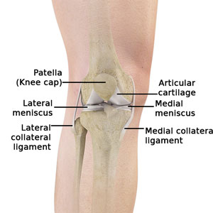
 Knee Joint Anatomy Bones Ligaments Muscles Tendons Function
Ligament Injuries To The Knee Johns Hopkins Medicine
 Normal Anatomy Of The Knee Joint Middletown Knee Treatment
 Knee Joint Anatomy Motion Knee Pain Explained
 Knee Joint Anatomy
 Clinical Anatomy Knee
 Anterior Anatomy Of Knee Joint Uptodate
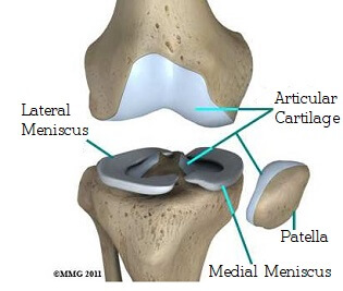
 Knee Anatomy The Knee Joint Cartilage
 4 Common Causes Of Knee Pain
 Anatomy Human Knee Joint Wall Mural
 The Knee Joint Articulations Movements Injuries
 Anterior And Posterior Aspects Of The Knee Netter
 Knee Joint Anatomy Bones Cartilages Muscles Ligaments
 Anatomy Of The Knee For Dancers Dance Work Balance
 Anatomy Of The Knee Joint Owlcation
 Anatomy Knee Joint Cross Section Showing The Major Parts Which
 The Knee Anatomy Injuries Treatment And Rehabilitation
 Knee Anatomy
 Knee Anatomy
Knee Joint Picture Image On Medicinenet Com
 Total Knee Replacement Orthoinfo Aaos


Belum ada Komentar untuk "Anatomy Of Knee Joint"
Posting Komentar