Anatomy Of The Knee Ligaments
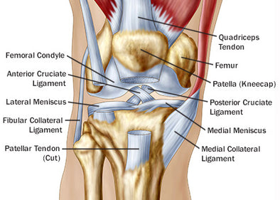
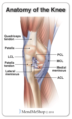
Another bone the patella kneecap is at the center of the knee. Knee ligament impose limitations on the movement of the knee allowing it to concentrate forces of the muscles on extension and flexion.
 Acl Solutions Acl Knee Anatomy And Diagram Images
Two concave pads of cartilage strong flexible tissue called menisci minimize the friction created at the meeting of the ends of the tibia and femur.

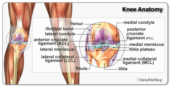
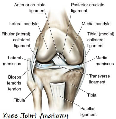
Anatomy of the knee ligaments. The anterior cruciate ligament and posterior cruciate ligament provide front and back anterior and. The knee is a hinge joint that is responsible for weight bearing and movement. Ligaments join the knee bones and provide stability to the knee.
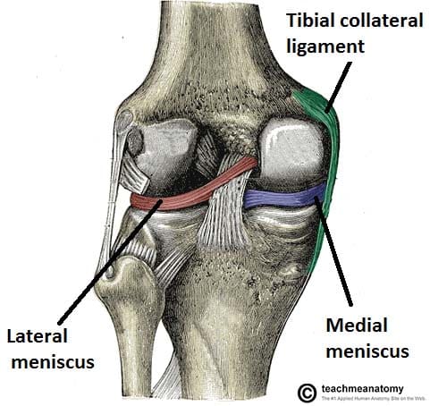
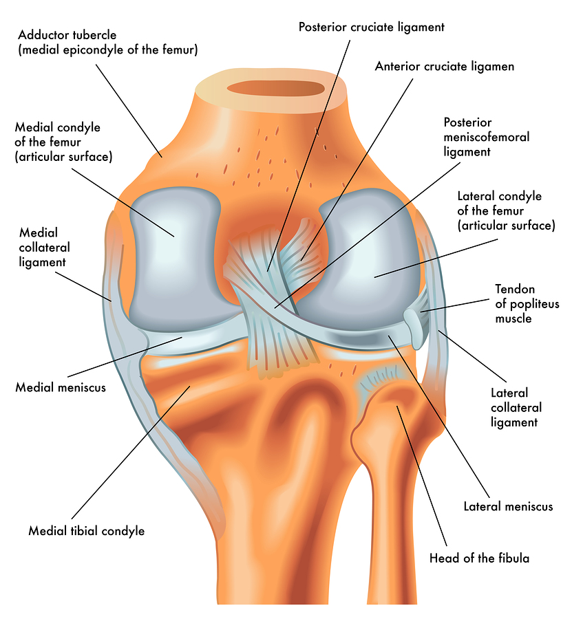
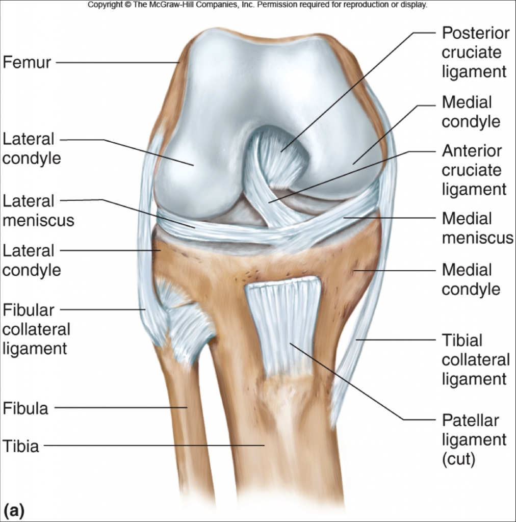
It consists of bones meniscus ligaments and tendons. The anterior cruciate ligament prevents the femur from sliding backward on the tibia or the tibia sliding forward on the femur. On the sides of the knee are the medial collateral ligament mcl and the lateral collateral ligament lcl.
These are called the cruciate ligaments and consist of the anterior cruciate ligament and the posterior cruciate ligament. The most common ligament injuries are acl tears mcl tears pcl tears and knee sprains which occur when the ligaments are overstretched. One ligament is on each side of the knee joint.
Femur the upper leg bone or thigh bone tibia the bone at the front of the lower leg or shin bone patella the thick triangular bone that sits over the other bones at the front of the knee or kneecap. The knee consists of three bones. There are also several key ligaments a type of fibrous connective tissue that connect these bones.
There is also a patellar ligament that attaches the kneecap to the tibia and aids in stability. The knee includes four important ligaments all of which connect the femur to the tibia. In knee joint anatomy they are the main stabilising structures of the knee acl pcl mcl and lcl preventing excessive movements and instability.
The medial collateral ligament on the inner side and the lateral collateral ligament on the outer side. There are four knee ligaments thick bands of tough tissue that serve to maintain the stability of the knee joint. Ligaments in the knee.
A belt of fascia called the iliotibial band runs along the outside of the leg from the hip down to the knee and helps limit the lateral movement of the knee. These two prevent sideways sliding of the knee joint ad also brace it against unusual movement. The largest joint in the body the knee moves like a hinge allowing you to sit squat walk or jump.
Ligaments of the knee. The knee is designed to fulfill a number of functions.
Knee Anatomy The Orthopedic Sports Medicine Institute In
 Knee Ligaments Joi Jacksonville Orthopaedic Institute
 Guide Knee Injuries In Bjj Fighters Market
 Knee Joint Picture Image On Medicinenet Com
 Royalty Free Knee Ligament Stock Images Photos Vectors
 Ligaments Of The Knee Knee Sports Orthobullets
 Knee Ligament Anatomy Animation

 Knee Joint Anatomy Bones Cartilages Muscles Ligaments
 14102 04b Tendons And Ligaments Of The Right Knee Anatomy
 Surgeons Discover Quirky Knee Ligament All Over Again
 Redding Hospital Knee Anatomy
 Why Are Women At Greater Risk For Acl Injuries

 The Knee Joint Articulations Movements Injuries
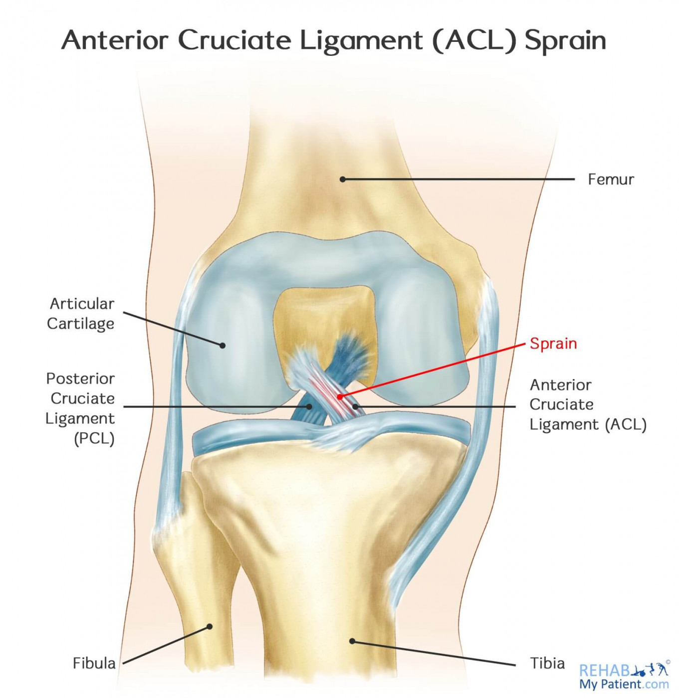 Knee Acl Sprain Rehab My Patient
![]() Knee Joint Anatomy And Function Kenhub
Knee Joint Anatomy And Function Kenhub
 Acl Vs Mcl Pcl Absolute Life Wellness Center
 Anterior Cruciate Ligament Acl Injuries Core Em
 Knee Pain The Center For Physical Rehabilitation
 Anatomy Of An Injury Acl Anterior Cruciate Ligament Tear
 Knee Pain Treatment Diagnosis Related Symptoms
 Knee Joint Anatomy Motion Knee Pain Explained
Common Knee Injuries Orthoinfo Aaos
Belum ada Komentar untuk "Anatomy Of The Knee Ligaments"
Posting Komentar