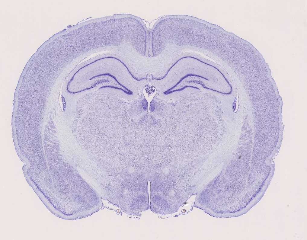
Anatomy Of Mouse Brain
Kovačević address correspondence to x. Rosen et al 2003 and it is this use that guided design of the atlas.
 Quantitative Rodent Brain Receptor Imaging Springerlink
Mouse brain gross anatomy atlas.
Anatomy of mouse brain. Interactive atlas viewer allen brain atlas. Dec 16th 1999 by. Under surface or right hind foot.
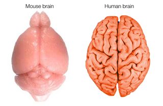
Separate the rest of the hippocampus from the cortex covering it along the surface of the hippocampus towards the ventral part of the hippocampus. The olfactory bulb volume takes about 2 of the mouse brain by volume in contrast to about 001 of the human brain. Mouse brain gross anatomy atlas.
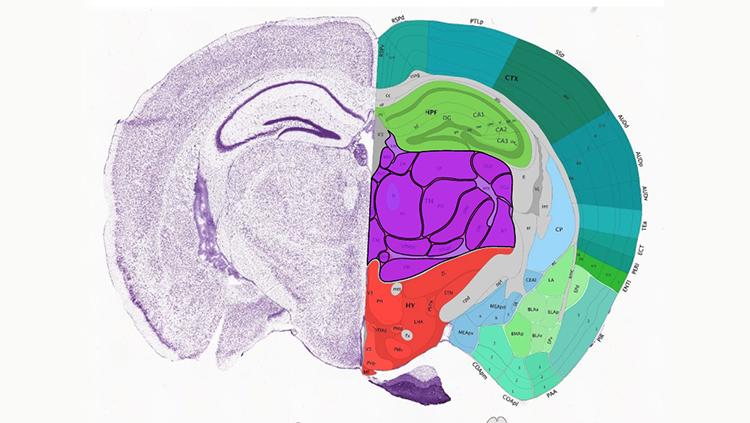
The allen mouse brain atlas includes a full color high resolution anatomic reference atlas accompanied by a systematic hierarchically organized taxonomy of mouse brain structures. Mbl is a research resource developed by the informatics center for mouse neurogenetics. Now you can see the dorsal part of the hippocampus.
Josette chen mouse imaging centre hospital for sick children 555 university avenue toronto ontario canada m5g 1x8. Edmundcape at hmsharvardedu code may be re used for non commercial use. Blood vessels of right hind limb.
The 2nd incision is about 15 2mm in front of the first one this incision you need cut into the lateral ventricle. The first incision is at the end of the hemisphere. We report here on the construction of the neuroterrain 3d mouse brain atlas nmba.
Edmund cape last updated. A three dimensional mri atlas of the mouse brain with estimates of the average and variability n. It was first presented as a poster and demo session at the incf booth of sfn 2009 in chicago.
Its main intended use is in conjunction with the mouse brain library mbl. Upper surface of right hind foot. Blood vessels of lower right hind limb.
The incision should be about 07mm deep for most adult mouse that you might not hurt the hippocampus while to expose it. The cerebral cortex of a mouse has around 8 14 million neurons while in those humans there are more than 10 15 billion. Skin removed from dorsal surface of right hind limb.
Superficial dissection of the neck and thorax. Development the scalable brain atlas is developed by rembrandt bakker in collaboration with many othersit uses exploratory work of gleb bezgin creator of the cocomac paxinos3d. In 2011 the reference atlas was updated to enable interactive online exploration of the atlas and to provide a deeper level of 3 d annotation for informatics analysis and viewing in the brain explorer 3 d viewer.
Blood vessels of axilla. Blood vessels of the neck. Now you need to free the hippocampus from the surrounding tissue.
 A Digital Atlas To Characterize The Mouse Brain Transcriptome

Untitled Document
A Digital Atlas To Characterize The Mouse Brain Transcriptome

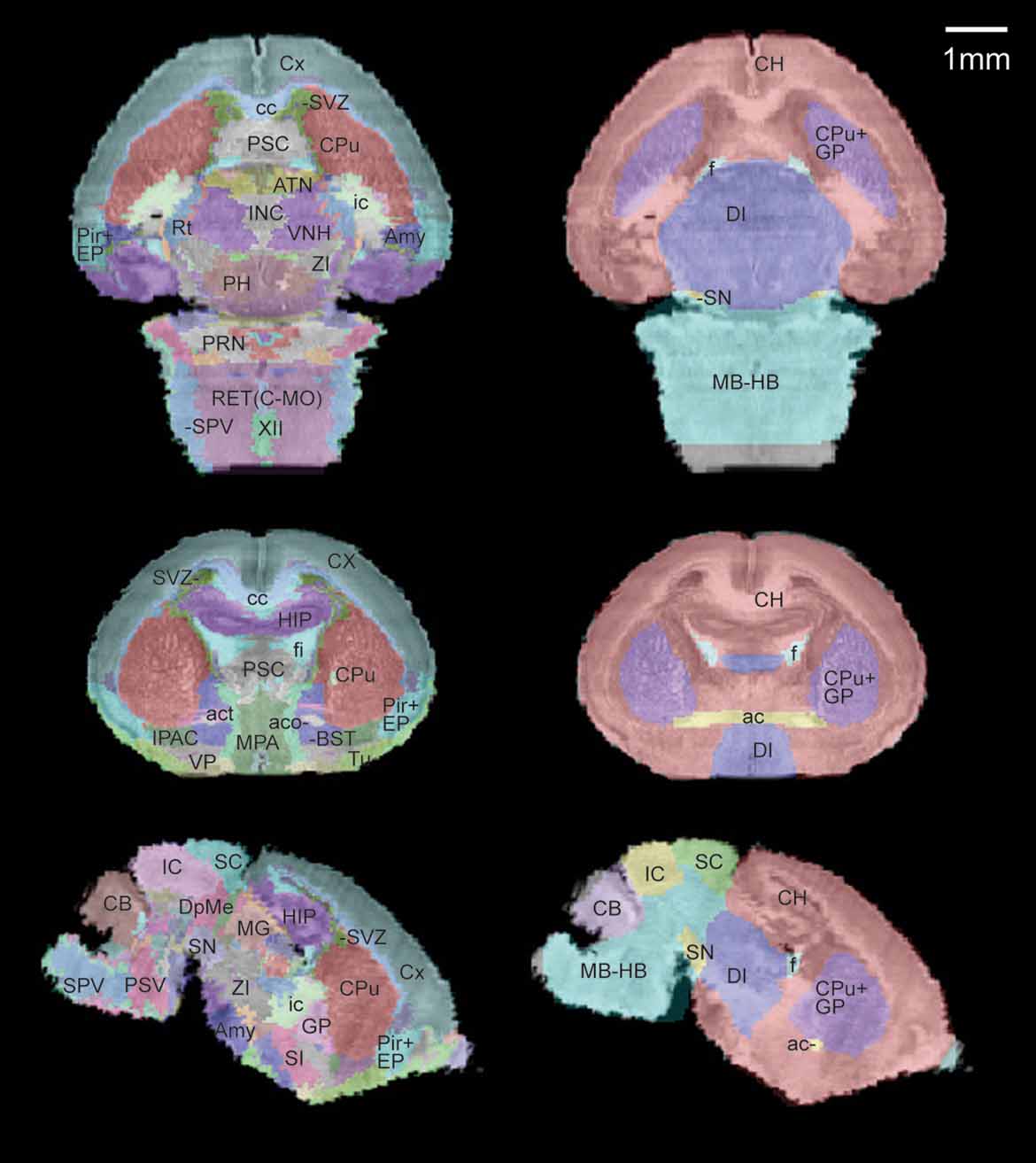
 Frontiers In Vivo 3d Digital Atlas Database Of The Adult
 1 Schematic Overview Of Basic Brain Anatomy In A Sagittal

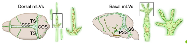 Anatomy News Flash Brain Drains Lymphatic Fluid Through Its
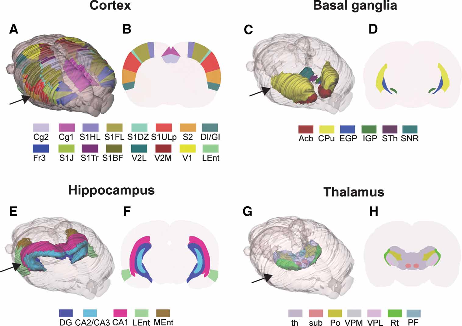
 Frontiers A High Resolution Anatomical Framework Of The
 Comparative Plasticity Of Brain Synapses In Inbred Mouse
 Comparative Anatomy Comparison Of The Brains Of The Adult
 Frontiers Three Dimensional Atlas System For Mouse And Rat
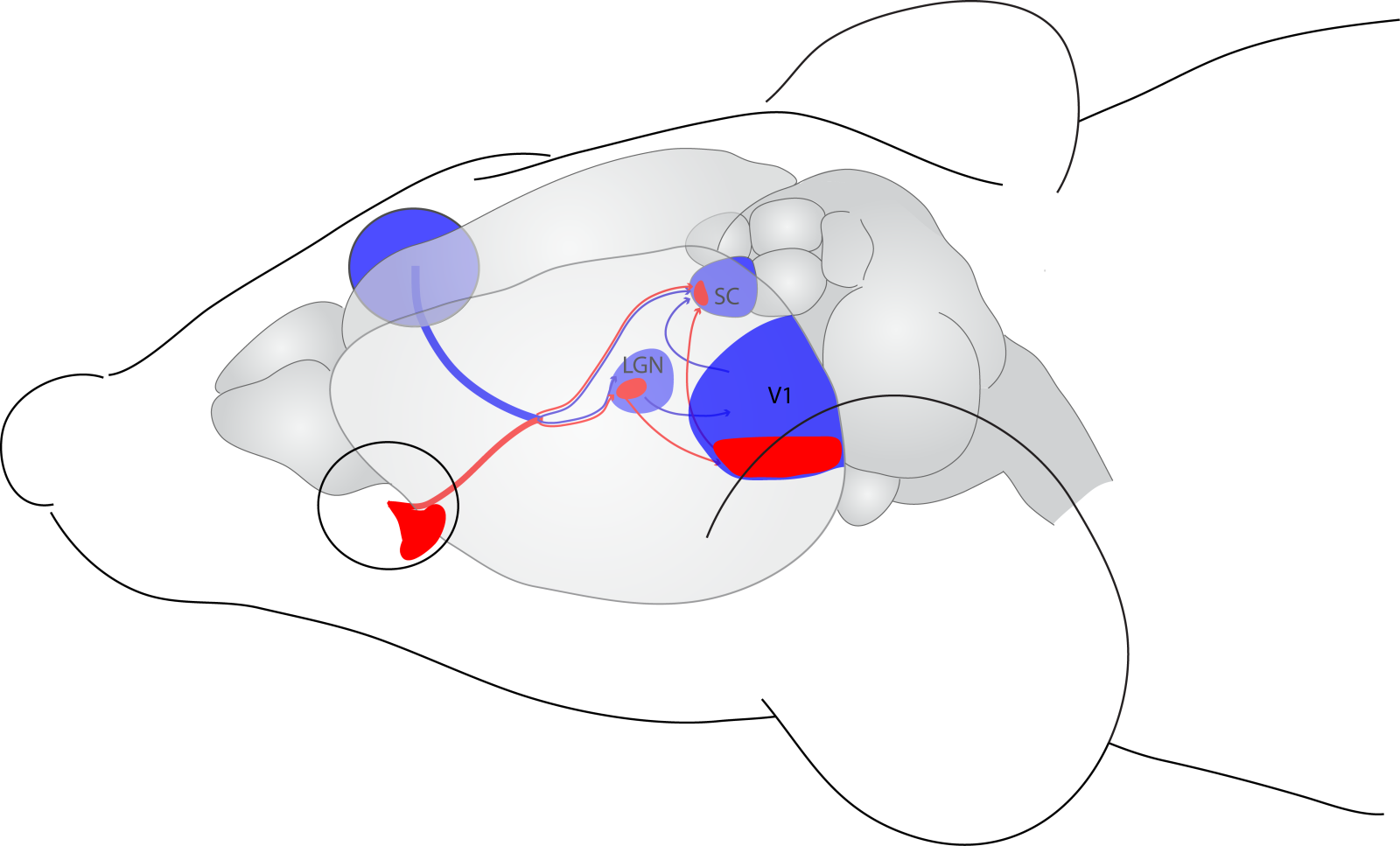 Seeing With Two Eyes Integration Of Binocular Retinal
 Brain Initiative
 How The Human Brain Gets Its Wrinkles Live Science
 Nervous System Sciencedirect
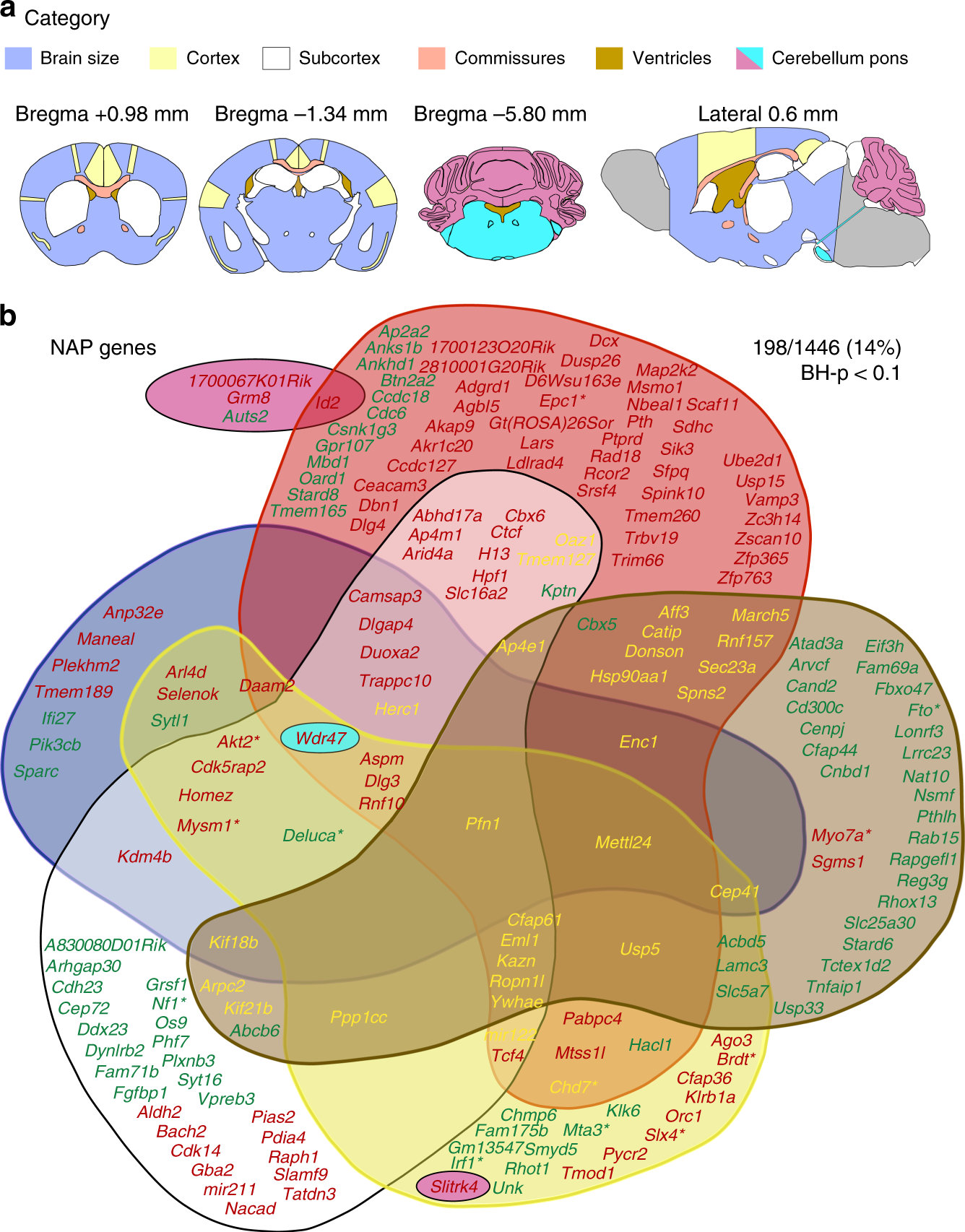 Large Scale Neuroanatomical Study Uncovers 198 Gene
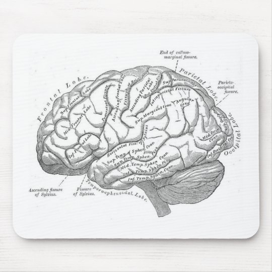 Vintage Brain Anatomy Mouse Pad
 Figure 2 From Genetic Dissection Of The Mouse Brain Using
Belum ada Komentar untuk "Anatomy Of Mouse Brain"
Posting Komentar