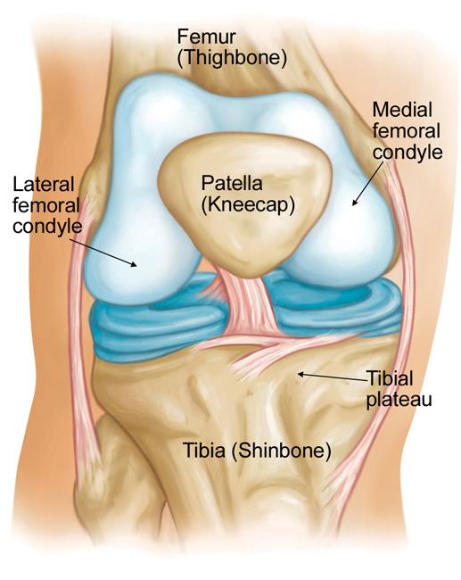
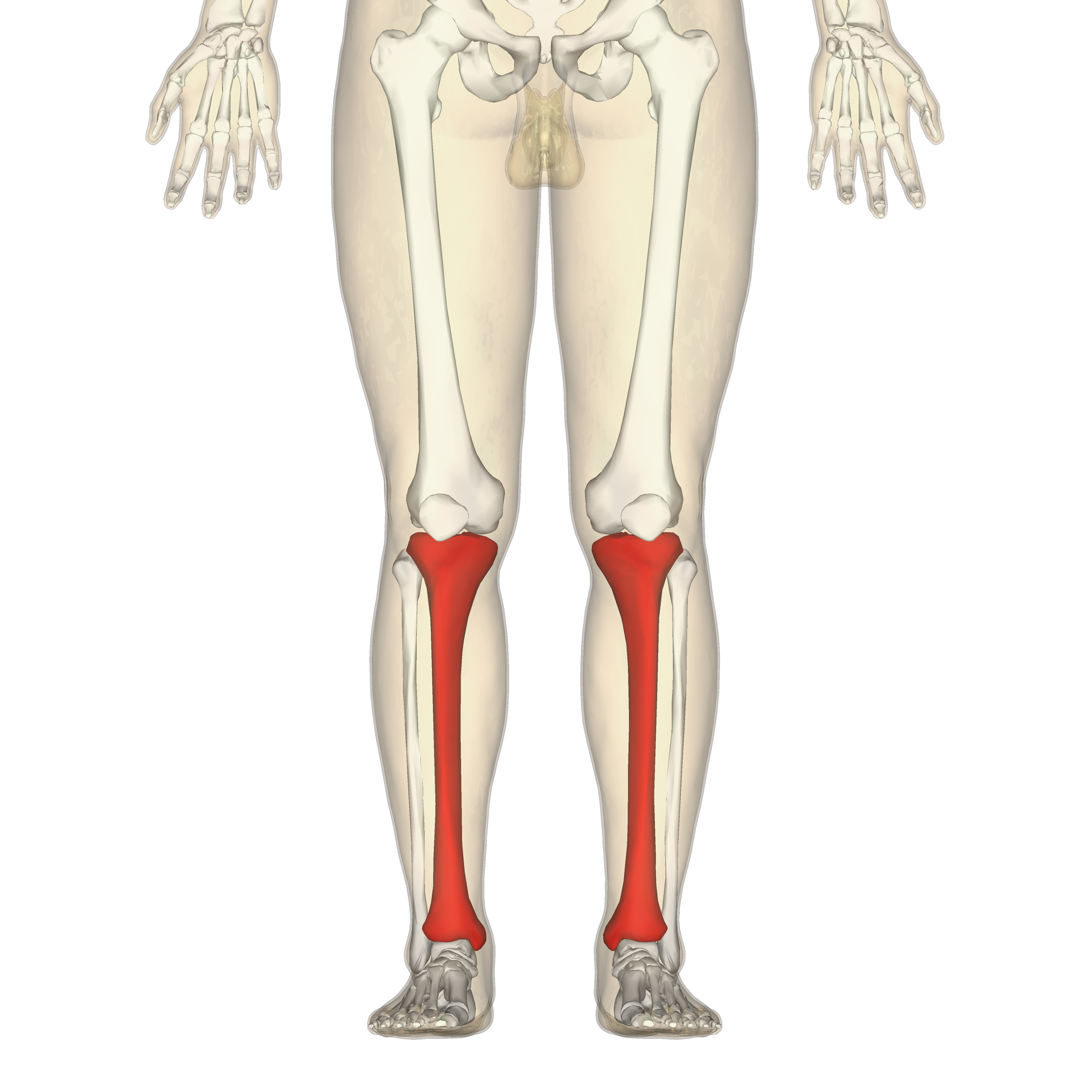
Tibial Plateau Anatomy
Fractures of the plateau affect knee alignment stability and motion. 83 this step off is lost when there is a posterior sag of the tibia associated with injury to the pcl and other secondary restraints to posterior translation of the tibia.
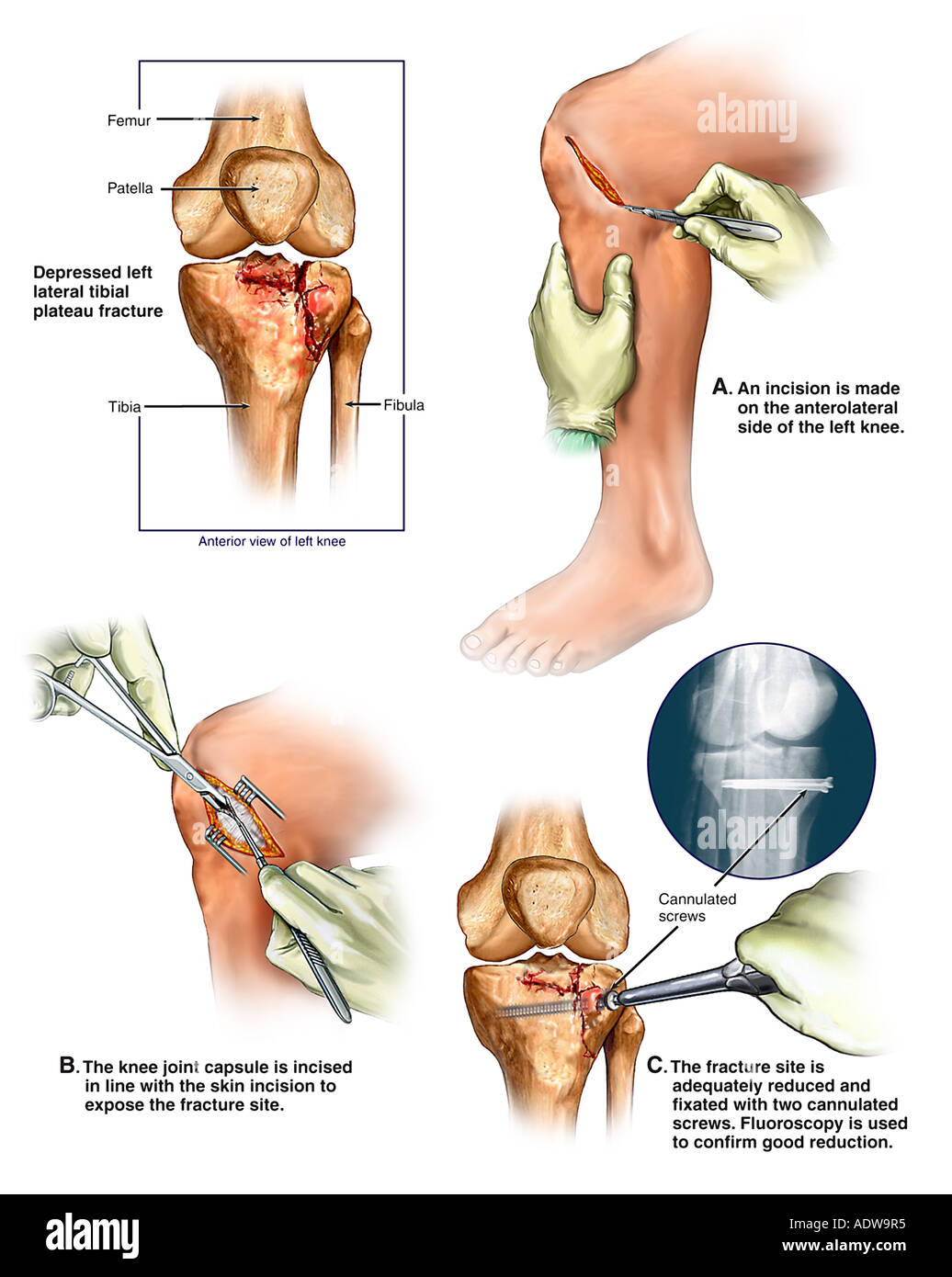
 Tibial Plateau Fractures With Subsequent Surgical Repair
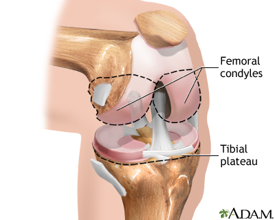
The condyles form a flat surface known as the tibial plateau.

Tibial plateau anatomy. The tibial plateau is made up of three osseous structures. Anatomy consist of medial and lateral plateau medial larger medial lower concave medial bone harder thus less likely to fracture lateral higher convex lateral cartilage thicker 3 vs. Lateral plateau is covered by meniscus tolerates incongruity better than medial plateau.
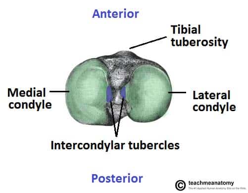
Located between the condyles is a region called the intercondylar eminence this projects upwards on either side as the medial and lateral intercondylar tubercles. Concave articular surfaces of the oval shaped medial and circular shaped lateral tibial condyles medial and lateral tibial plateaus. A fall from a height or a hit to the thigh can drive the femur.
Medial plateau larger than lateral medial is concave in sagittal plane golf tee lateral is convex more proximal golf ball creates 3 o of varus proximal tibia. Lateral plateau more commonly fractures. 4 mm lateral medial.
The lateral plateau is smaller as well as convex while the medial plateau is larger and slightly concave. The tibial plateau is one of the most critical loadbearing areas in the human body. This structure articulates with the femoral condyles to form the key articulation of the knee joint.
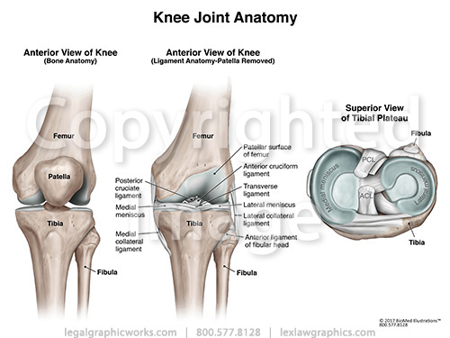
The tibial plateau is composed of two parts. Early detection and appropriate treatment of these fractures are critical for minimizing patient disability and reducing the risk of documented complications particularly posttraumatic arthritis. Normal posterior slope 10 o.
Central non articular intercondylar area. Normally the medial tibial plateau protrudes anteriorly 1 cm beyond the medial femoral condyle when the knee is flexed to 90 figure 20 20. The tibial plateau is an important weight bearing part of the body that connects the thighbone femur to the shinbone via ligaments.
The lateral plateau the medial plateau along with the intercondylar eminence.
 Tibial Plateau Fractures Physiopedia
 Pdf Anatomical Morphometry Of The Tibial Plateau In South
 Tibial Plateau Fractures
 Osteonecrosis Of The Knee Orthoinfo Aaos
 Comminuted Proximal Tibia Fracture Of Tibial Plateau Stock
 What Is Fractured Tibial Plateau Or Tibial Plateau Fracture
 Sports Docs Weekly Blitz Tibial Plateau Fracture Ends Jj
Fractures Of The Proximal Tibia Shinbone Orthoinfo Aaos
 Tibial Plateau Stock Photos Tibial Plateau Stock Images
 Superior View Tibial Plateau Medical Illustration Human
 Tibial Plateau Fractures Trauma Orthobullets
 Broken Knee Knee Fracture
 The Tibia Proximal Shaft Distal Teachmeanatomy
 Anterior Tibial View Of Knee Joint Anatomy
 Proximal Tbial Anatomy
 What Is Tibia Bone Location Function Anatomy Pictures
 Tibial Plateau Fracture Schatzker Type V Radiology Case
 Tibia Wikipedia
 X Knee Startradiology
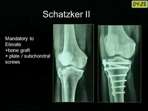
 Revisiting The Schatzker Classification Of Tibial Plateau
 What Is Fractured Tibial Plateau Or Tibial Plateau Fracture
 Tibial Plateau Fractures
 Tibial Plateau Fractures Musculoskeletal Medicine For
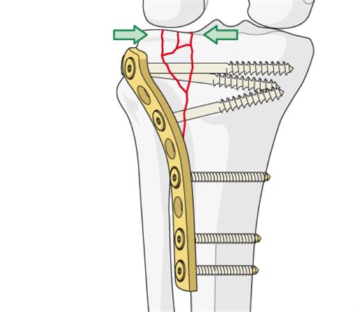
 Proximal Tibia Reduction Fixation Orif Plates
 Medical Exhibits Demonstrative Aids Illustrations And Models
The Anatomy Of The Articular Surface Of Kangaroo Tibial
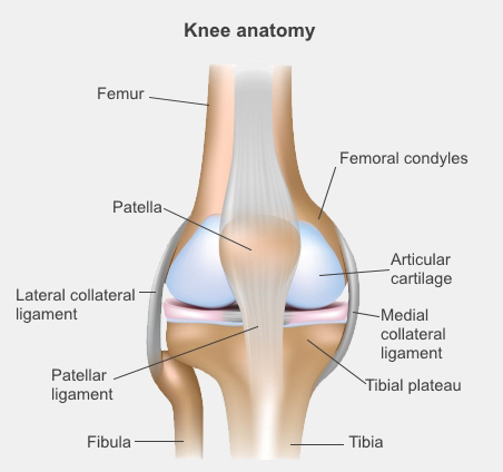
 Anatomy Knee Restoration Center Of Indiana
 Tibial Plateau Fractures Background Anatomy Pathophysiology
 Tibial Plateau Fracture Wikipedia
 Knee Joint Replacement Series Normal Anatomy Medlineplus
 Tibial Plateau Fracture Radiology Reference Article


Belum ada Komentar untuk "Tibial Plateau Anatomy"
Posting Komentar