Anatomy Pictures Of The Stomach
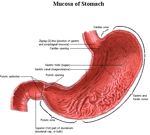
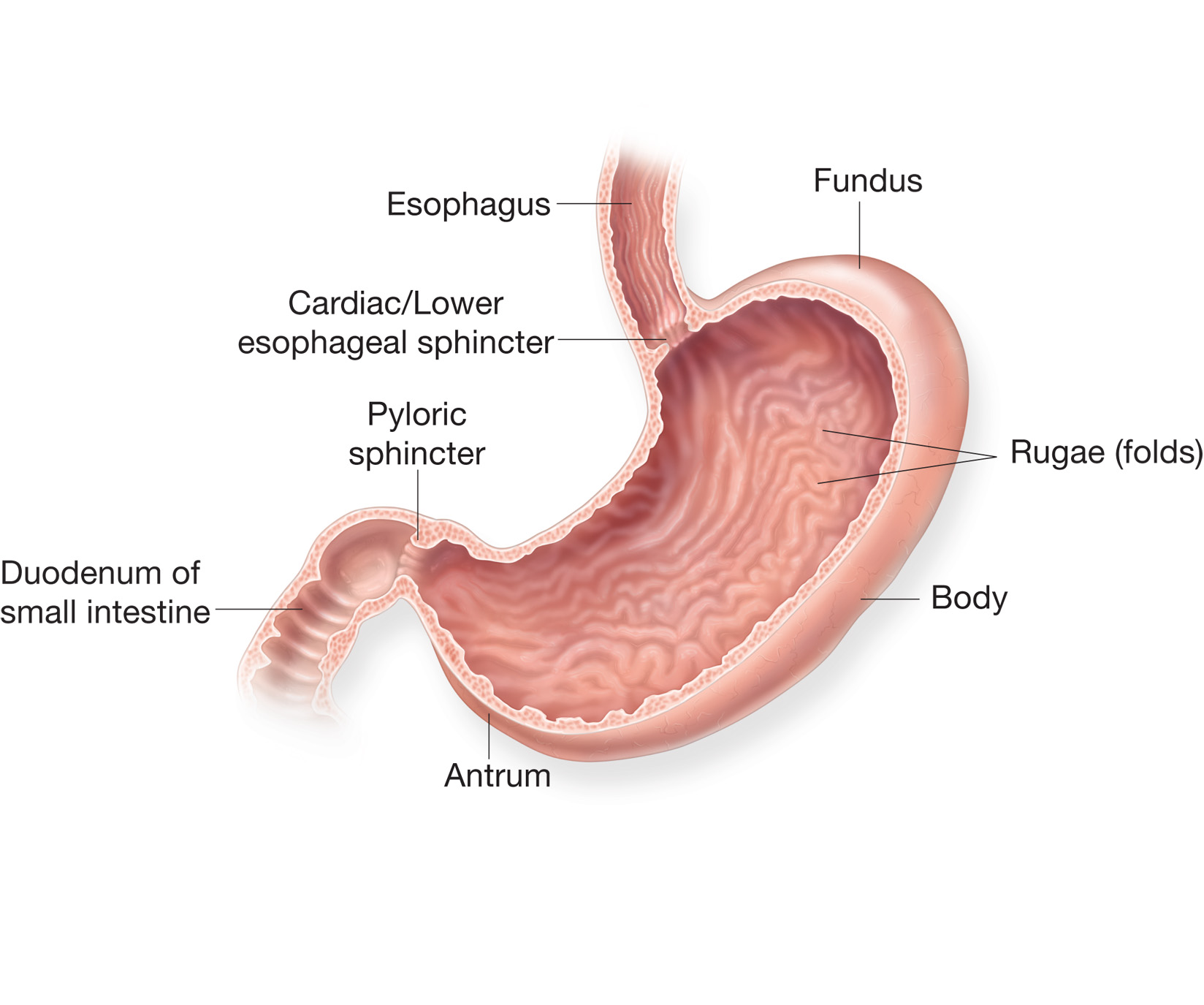
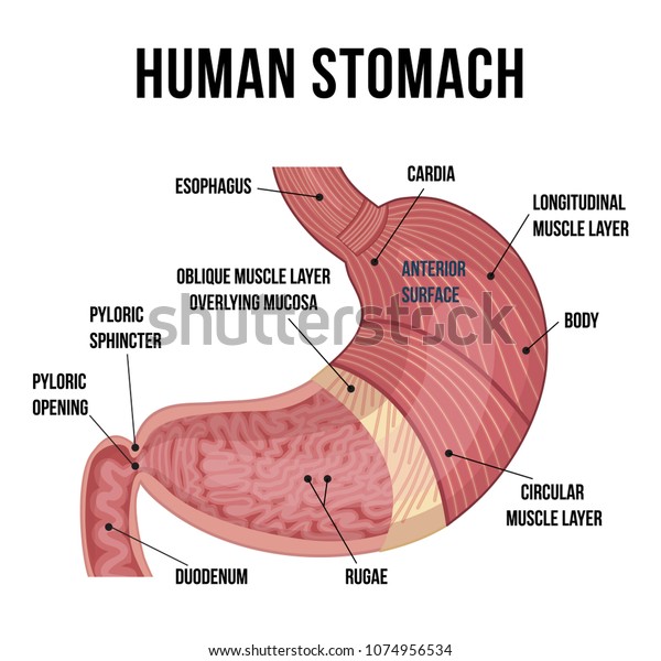
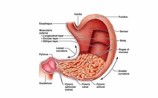
The cardia fundus body and pylorus. Picture of the stomach.
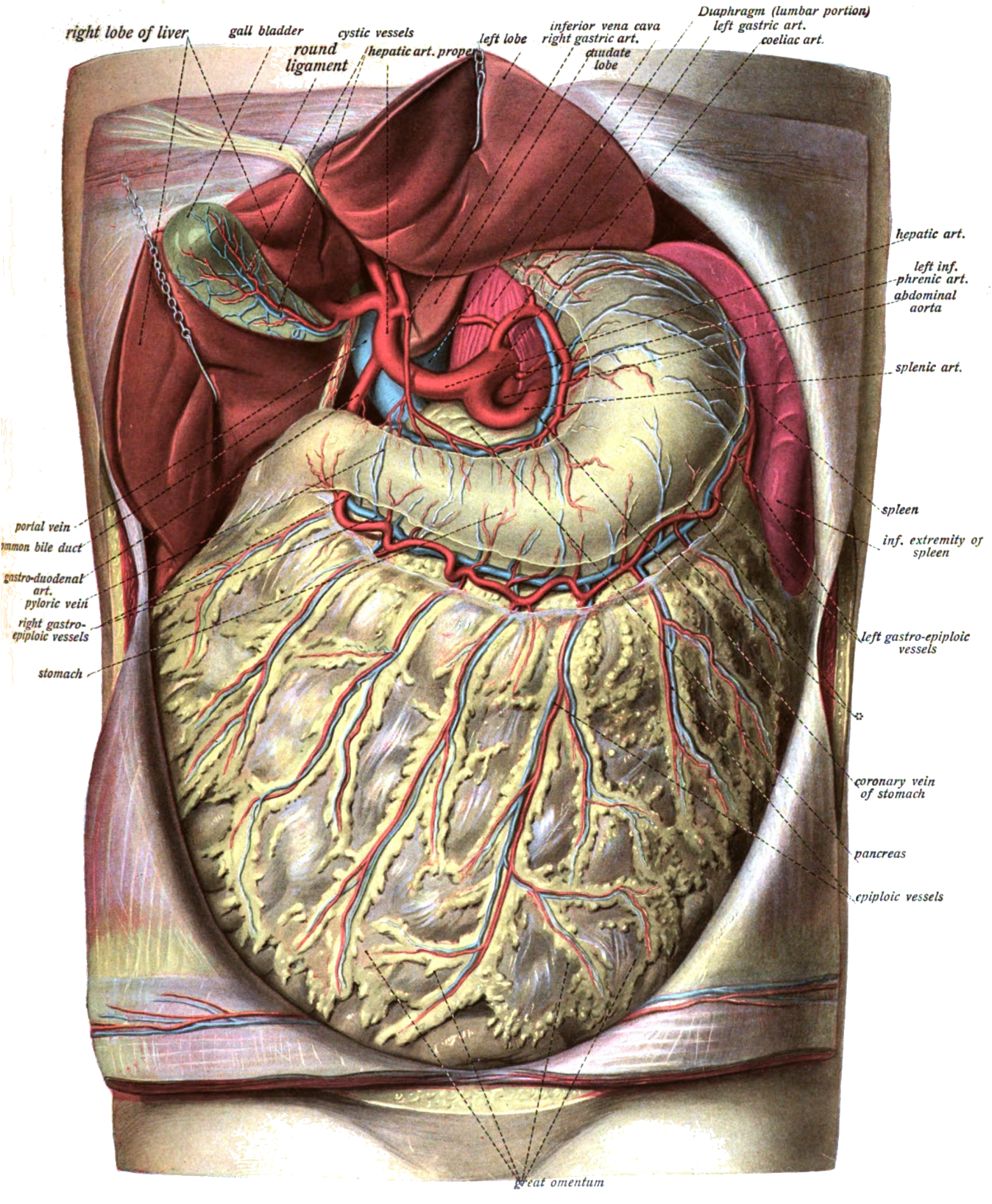
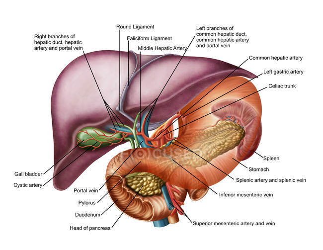 Anatomy Of Liver With Stomach And Pancreas Digestive
These include parietal cells chief cells mucous neck cells and enteroendocrine cells.

Anatomy pictures of the stomach. The stomach releases enzymes and acid to help digest food. If the lining of the stomach is examined with a hand lens one can see that it is covered with numerous small holes. Learn about its function parts abdominal conditions and more.
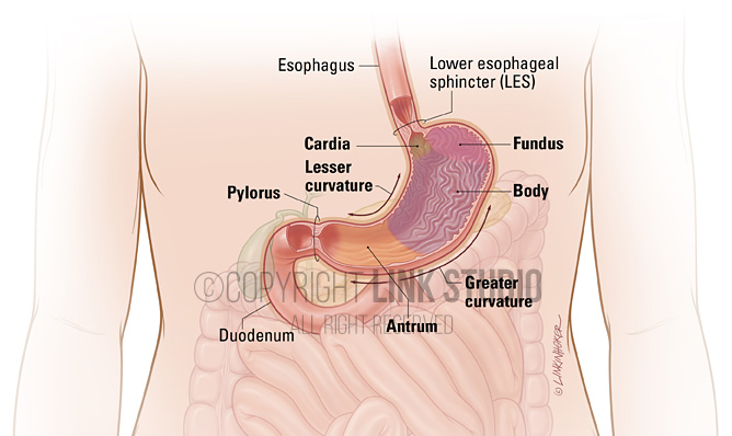
The stomach receives food from the esophagus. The stomach has four main anatomical divisions. Stomach anatomy and its parts.
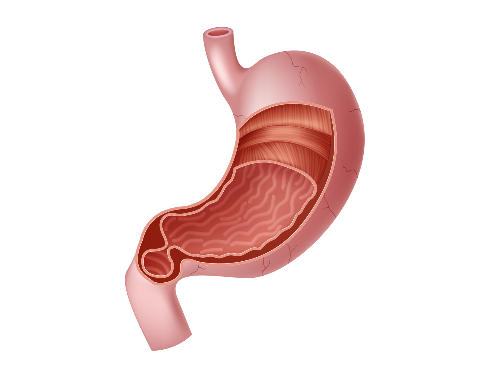
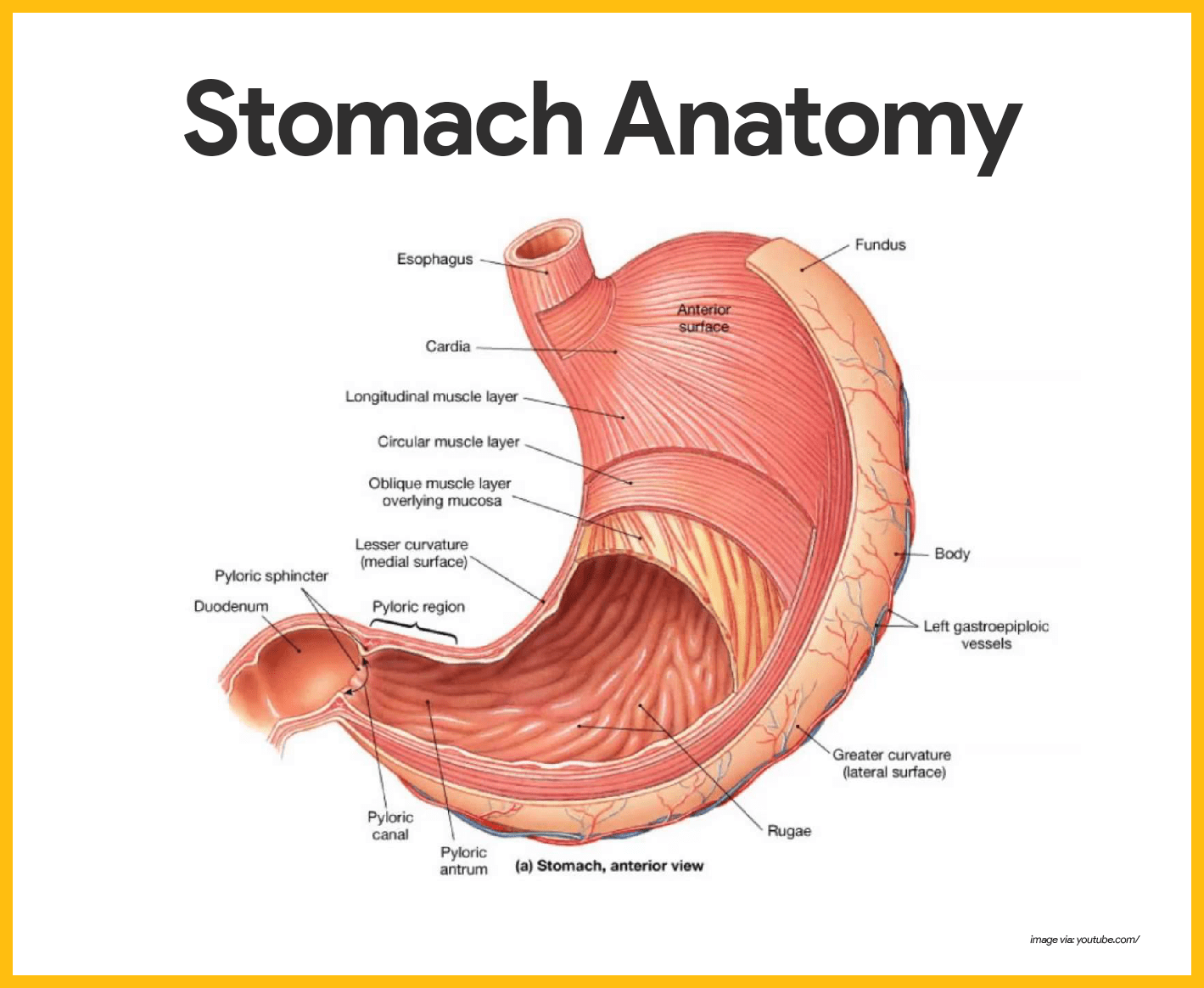
Fundus the rounded often gas filled portion superior to and left of the cardia. It is about 6 meters 20 feet long and extends from the pylorus of the stomach to the ileocecal junction. Ridges of muscle tissue called rugae line the stomach.
Location of the intestines. When that muscle doesnt relax properly it feels like it is difficult to swallow. Body the large central portion inferior to the fundus.
The stomach contracts and churns turning stomach contents into a partially digested substance called chyme. These are the openings of gastric pits which extend into the mucosa as straight and branched tubules forming gastric glands. The image above shows rugae on the surface of a dogs stomach.
On the opposite end of the stomach the pyloric sphincter regulates the speed at which food moves down to the small intestine. The small intestine is divided into. Anatomy of the stomach area several key organs are packed closely with the stomach in the abdominal cavity including the liver whose smaller left lobe is located superior to the stomach and whose large right lobe occupies the same space in the upper right quadrant as the stomach and left lobe combined.
The stomach secretes acid and enzymes that digest food. The stomach lining contains muscular folds called rugae. Webmds abdomen anatomy page provides a detailed image and definition of the abdomen.
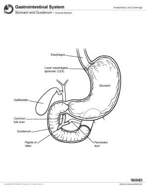
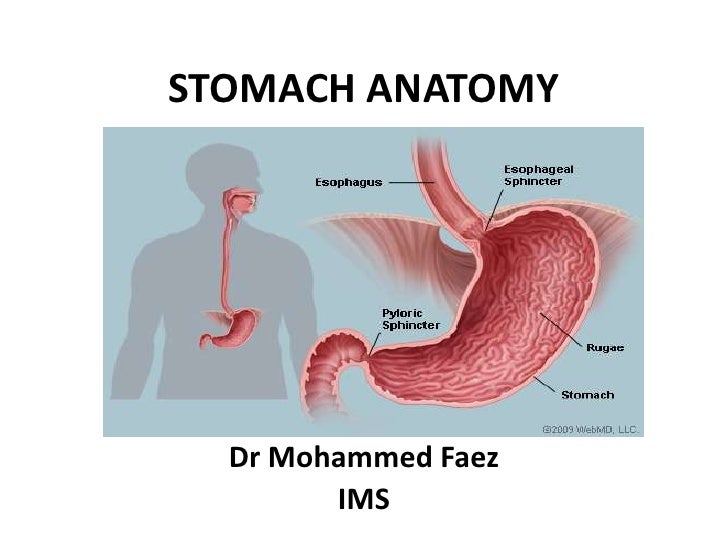
Picture of abdomen the abdominal cavity is the part of the body that houses the stomach liver pancreas kidneys gallbladder spleen and the large and small intestines. As food reaches the end of the esophagus it enters the stomach through a muscular valve called the lower esophageal sphincter. Anatomy of the stomach.
Cardia surrounds the superior opening of the stomach at the t11 level. The human intestine consists of two segments. The diaphragm marks the top of the abdomen and the horizontal line at the level of the top of the pelvis marks the bottom.
Divisions of small intestine. The much larger glands of the fundus and body of the stomach the site of most chemical digestion produce most of the gastric secretions. These glands are made up of a variety of secretory cells.
The esophageal sphincter separates the esophagus and the stomach. The separation between the esophagus and the stomach is a valve called the lower esophageal sphincter.
 Human Stomach Anatomy Diagram Human Anatomy Body Picture
 In Human Anatomy Does The Stomach Touch The Heart Quora
 Anatomy For Radiology Abdomen Glass Box
 Stomach Cross Sectional Anatomy
 Stock Illustration
 Stomach Anatomy
 Human Digestive System Anatomical Poster Physiology Chart Of Oral Cavity Glands Stomach Liver Pancreas And Duodenum Silk Size 24 Width X 32
 Understanding The Human Stomach Anatomy With Labeled
![]() Stomach Anatomy Function Blood Supply And Innervation
Stomach Anatomy Function Blood Supply And Innervation
 Nicoleshenting Human Anatomy Stomach System Art Silk Poster 13x20 24x36 Inch Body Map Pictures For Medical Education 011
 The Stomach Structure Of The Stomach Anatomy Of The
 Gastrointestinal Tract 2 The Structure And Function Of The
 Stomach Anatomy Overview Gross Anatomy Microscopic Anatomy
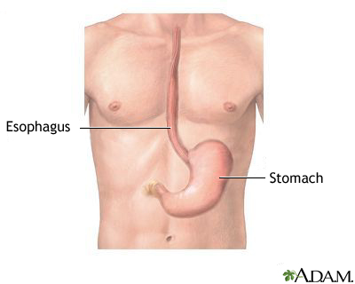 Esophagus And Stomach Anatomy Medlineplus Medical
![]() Stomach Anatomy Function Blood Supply And Innervation
Stomach Anatomy Function Blood Supply And Innervation
 Horse Stomach Model
 Digestive System Anatomy Link Studio
 Anatomy Of The Human Healthy And Unhealthy Stomach
 Stomach Anatomy By Tigatelu On Dribbble
 Digestive System Anatomy And Physiology Nurseslabs
 The Anatomy Of The Abdomen Human Stomach Health Life Media
 Brainmedico
 Gross Anatomy Of The Stomach Diagram Quizlet
/male-stomach-layers-anatomy--illustration-758312869-5a00caa49e9427003ca76fdc.jpg) Illustrated Anatomy Of The Stomach
Illustrated Anatomy Of The Stomach
 Greater Omentum Wikipedia
![]() Human Internal Organs By Svitlana Babych
Human Internal Organs By Svitlana Babych
 Human Stomach Anatomy Vector Illustration Stock Vector
![]() Stomach Anatomy Function Blood Supply And Innervation
Stomach Anatomy Function Blood Supply And Innervation
 Stomach Diaphragm Esophagus Fetal Pig Anatomy Lab
 Ch23 Stomach Anatomy
 1 Stomach


Belum ada Komentar untuk "Anatomy Pictures Of The Stomach"
Posting Komentar