Female Anatomy In Pregnancy
It has three sections. The uterus is a hollow pear shaped organ that is the home to a developing fetus.
 Vintage Print Of Female Internal Anatomy During Pregnancy
When you are between 39 and 41 weeks your pregnancy is considered full term and your baby is ready to be born.
Female anatomy in pregnancy. Body and beauty 4636845 views. Demystifying female anatomy is key to good sexual functioning whether youre a mature experienced adult or looking to learn about womens sexual organs for the first time. Inside the uterus or womb is a hollow pear shaped organ.
It starts at week 28 of your pregnancy and ends with the birth of your baby. This sonogram is used to determine fetal anomalies the babys size and weight and also to measure growth to ensure that the fetus is developing properly. 9 big mistakes every woman should avoid during pregnancy duration.
The portion of the uterus superior to the opening of the uterine tubes is called the fundus. Vagina the vagina is a tube that connects your vulva with your cervix and uterus. Everything from belly size to heartbeat speed will change over the 9 months leading up to childbirth.
A full term fetus non morphing is included with the model that is scaled to fit inside the expanded uterus. What is an anatomy ultrasound. Its what babies and menstrual blood leave the body through.
Its also where some people put penises fingers sex toys menstrual cups andor tampons. Pregnancy is a time of great physical and emotional change for women. The internal reproductive organs in the female include.
Understanding womens sexual or reproductive organs such as the vagina uterus and vulva is as integral to sex as understanding the penis. The cervix connects the vagina to the uterus and produces mucus. Lets focus on the third trimester.
The package includes the skin of the female and the uterus showing the changes in the lower abdominal and pelvic region that take place during pregnancy. When the pregnancy hits the 20th week of gestation an anatomy ultrasound is often ordered. The internal parts of female sexual anatomy or whats typically referred to as female include.
It also is known as the birth canal. During these final weeks your baby continues to grow and develop. The ovaries are glands which produce female sex hormones and egg cells ova.
The human vagina and other female anatomy. The vagina is a canal that joins the cervix the lower part of uterus to the outside of the body. The model can be purchased with or without texture maps for the female skin and hair.
Its average size is approximately 5 cm wide by 7 cm long approximately 2 in by 3 in when a female is not pregnant. Two fallopian tubes one on each side stretch from the ovaries to the uterus.
Can A Man Become Pregnant Carry And Give Birth To A Baby
 Female Pelvic Anatomy Early In First Pregnancy Medical Exhibit
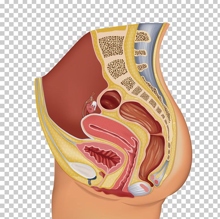 Female Reproductive System Organ Human Reproductive System
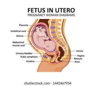 Female Pregnant Anatomy Images Stock Photos Vectors
 Vialibri Anatomy And Physiology Of The Female Generative
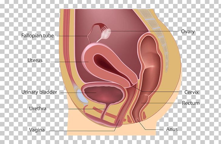 Retroverted Uterus Anatomy Vagina Pelvic Floor Png Clipart
The Amazing Female Pelvis Designed For Giving Birth
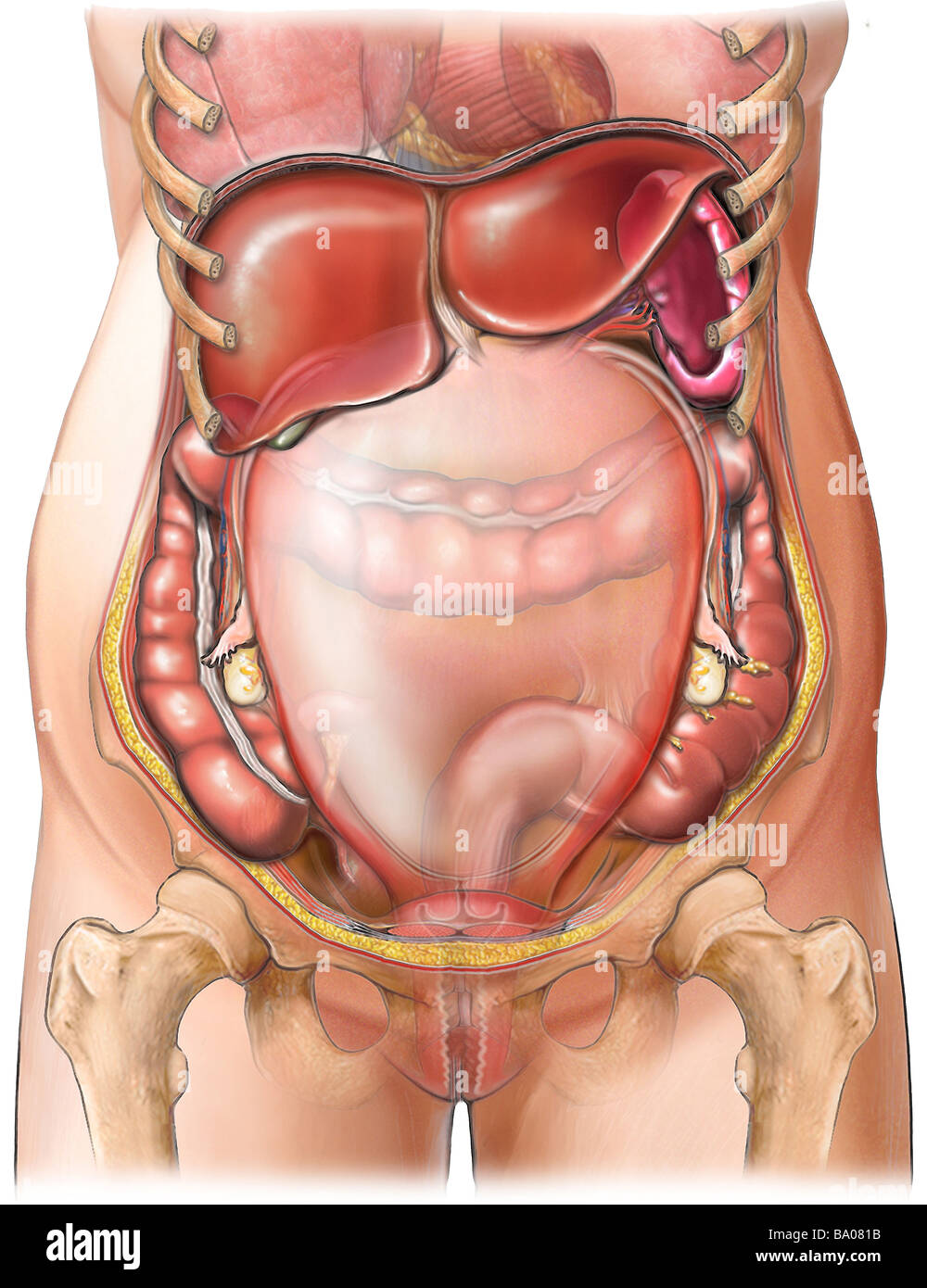 This Medical Illustration Depicts The Anatomy Of An Adult
 Female Reproductive Anatomy Reproductive Medbullets Step 1
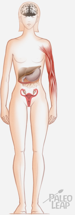 A Paleo Guide To Pregnancy Paleo Leap
 Human Female Pelvic Section Pregnancy Anatomical Model
 Female Pelvis With Uterus In The Ninth Month Pregnancy Model Female Anatomy Pelvis With Baby Model Buy Female Pelvis With Uterus Female Anatomy
 Progesterone Hormone Britannica
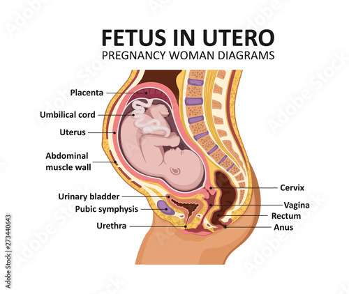 Fetus In Utero Pregnancy Women Diagrams Pregnant Female
 Budget Pregnancy Pelvis Model Gjptpq
 Female Reproductive System Purpose 1 Produce Estrogen And
 Usd 65 55 4d Master Human Body Assembly Model Organ Anatomy
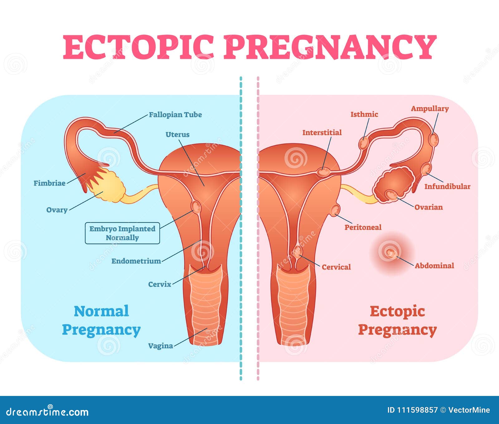
 Ectopic Pregnancy Or Tubal Pregnancy Medical Diagram With

 The Female Anatomy Freedom Products Juju

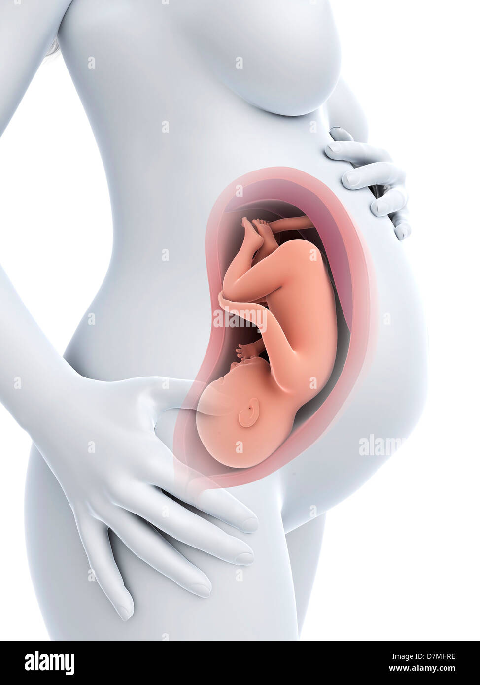 Female Anatomy Pregnancy Stock Photos Female Anatomy


Belum ada Komentar untuk "Female Anatomy In Pregnancy"
Posting Komentar